Foot and mouth
disease is an economically devastating and a highly contagious disease of
cloven hoofed animals as a result of loss of production; cost of treatments and
it is the main barrier to trade of animal and animal products at local and
international markets (James and Rushton, 2002). The
disease is characterized by low mortality in adult cattle and massive
mortality in young calves due to acute Myocarditis (kandeil
et al., 2013).
It is a virus of the genus Aphtho-virus in the
family of Picronaviridae of which seven
immunologically distinct serotypes (O,
SAT2, SAT1, A, C, Asia 1 and SAT 3) are circulating in the wide field but
multiple subtype strains have evolved within each serotypes. This variation
causes failure of the prevention and control options as infection or
immunization with one serotype doesn’t confer protection against other
serotypes and fails to protect other subtypes of the same strains (Paton et al., 2005).
FMD outbreaks have been reported in Ethiopia and the situation indicated
that, four serotypes (A, O, C and SAT 2) have been identified (Sahle,
2004). According to (Ayelet et al., 2009) study, serotype O
(73.30%) was the most prevalent followed by A (19.50%), SAT-2 (4.10%), SAT-1
(1.80%) and C (1.40%) during outbreaks. Besides this, Klein.
2009) indicated that serotype O is the most prevalent strain worldwide. The incident of new strains and uncontrolled movement
are some of the risk factors of FMD outbreak in Ethiopia (Bewket
et al., 2012). However, a
study on the specific serotype of virus that causes outbreak is limited. So, identification
of the virus strain from outbreak is very important to apply emergency vaccines
(Yang et al., 2013). Outbreak
based study is also important in the disease endemic areas to detect newly
emerging variants despite the fact the existence of other serotypes or
new virus subtypes might
cause outbreak. Hence, generation
of updated information on genetic
characteristics of this strain could
help to understand the circulating strains and institute disease prevention and
control packages. Therefore, the objectives of this outbreak study were: to
identify serotype O virus from clinical and asymptomatic Foot and mouth disease
cases in the dairy farm and to genetically characterize the virus from farm
outbreaks.
MATERIALS AND METHODS
Farm based outbreak
study
Foot and mouth disease outbreak had been
occurring in Welmera district Menagesha
kolobo kebele in private
dairy farm by mid-September 2016/2017. Welmera
district is a part of Oromiya special zone
Surrounding Finfinne and the district is 40 km away
from the capital city, Addis Ababa. The area is situated at 90- 04'- 9o 13' N
latitude and 38o 29'-38o 39' E longitude. It is bordered on the south by the Sebeta Hawas, on the west by West
shewa zone, on the North by Mulo
district, on the Northeast by the Sululta. Menagesha Kolobo and Holeta are the towns located in the district. The average
altitude of the area ranges from 2200-2500 meter above sea level and the the area is characterized by short rainy pattern from
February to April and a long rainy season from mid-June to September. The
annual temperature and rainfall ranges from 18°C to 24°C and 1000 to 1100 mm,
respectively (CSA, 2009 )
FMD Infected cows
reared by producers
Cattle
that had manifested clinical signs of the disease and those with asymptomatic
cases in the farm were included in the study. Cattle determinants such as age,
sex, breed and body conditions were considered during sampling.
Sampling
techniques and Ethical consideration
Clinically and acutely sick cattle were
physically examined in the affected farm. Moreover, clinical parameters and
clinical signs were recorded so as to know the status of infection in the
farm. Further, clinically sick and
acutely infected animals were purposively screened and ten cows were considered
for clinical diagnosis and laboratory studies. Ethical Consideration was also
used to alleviate suffering of
animals from sampling during sample collection.
Sample
collection from clinical and asymptomatic cases
Samples were collected from clinically sick
cattle and from those which had healing lesion in the mouth, dental pad or on
the feet and the asymptomatic cows in the farm (Kafeero
et al., 2016). Active epithelium were
aseptically collected from gum and tongue of affected cows and put in a bottle
with transport medium having glycerol and 0.04M phosphate buffer saline (PBS)
with some antibiotics and antifungal drugs.
Probang samples were also taken from foot and
mouth disease infected and asymptomatic cattle (OIE, 2004).
Foot and
Mouth disease virus isolation and characterization in cell culture
The samples were processed and cultured on
BHK-21 cell monolayer with three subsequent passages. About 1 gram of tissue
was washed three times using sterile phosphate buffered saline containing
antibiotics and antifungal drugs on petridish and
transferred to sterile mortar. The minced tissues were homogenized in sterile
sand with pestle and mortal. Nine ml of PBS was added to the homogenized
tissues and five percent antibiotics were added to make it ten times as that of
the epithelial tissue in order to produce ten percent suspension (OIE, 2012).
Then, the suspension was transferred to test tube and clarified by
centrifugation at 3500 rpm and the supernatant was inoculated to baby hamster
kidney- 21 cells and incubated at 370 c with 5% CO2. The cells were
monitored daily to see the formation of virus induced cythopathic
effects (CPE). The virus is cytocidal and CPE is
characterized by a fast destruction of the cell monolayer and infected cells
were disrupted and detached from the flask. When CPE appreciated in the
infected cases, supernatants of the clinical tissues had been typed by antigen
detection ELISA as per the recommended procedure (Bhattacharya et al., 1996). If no CPE was observed
following three blind passages, the virus isolation was considered as negative
(OIE, 2012).
Detection
and subtyping of O serotype from field based outbreak cases
The foot and mouth disease virus serotyping
was performed by antigen detection sandwich ELISA with appropriate combinations
of antiFMDV monoclonal antibodies (MAbs), used as coated and conjugated antibodies. The test
was done for typing of FMD viruses. The kit was made for detecting and typing
of FMD viruses serotypes such as type O, A, SAT 1 and SAT 2. A pan FMD test, detecting any isolates of
serotypes O, A, C, Asia 1 and some of the SAT serotypes were incorporated in
the kit to complement the specific typing and to detect foot and mouth disease
viruses which could have escaped binding to selected serotype-specific MAb. The micro plates were covered with catching MAbs. The test was done as per the manufacturer’s
procedure and six samples were tested on
a microplate containing 96 wells, one positive
control for each FMD types O,A, SAT1 and SAT2 and negative controls were
included in each plate. These controls were already incorporated into the ELISA
microplate trapped by the respective catching MAb. First samples were diluted half in diluent buffer and
50μl of each diluted sample was distributed in 72 wells of A-F rows and
two replicates of each-specific catching MAb and for
the pan-FMDV MAb. Then, 50μl of diluents per
well were added in all wells of G and H rows (positive and negative control,
respectively), then plates were incubated at 25o C for 1hour. After incubation,
all fluids on the plates were discarded and the remaining residual fluids were
removed. Then 200μl of washing solution were added and incubated for 3min
at room temperature, subsequently wells were emptied and the washing repeated
twice. Then the residual fluids were removed by tapping on clean absorbent
paper and 50μl of conjugate A was added from columns 1 to 8 and the same
volume of conjugate B was added from columns 9 to 12. Plates were covered and
incubated at room temperature for 1hour. After incubation, 50μl of
substrate per well was added to all wells and plates were covered and left at
room temperature for 20minutes in the dark. The reaction was stopped by adding
50μl of stop solution. Immediately after stopping, reading the optical
density (OD) of each well was done at 450 nm wavelength using micro plate
reader. Criteria for test validity: The positive controls were expected to give
OD values of 1.0 unit or higher in the type-specific reactions and in the
pan-FMDV reaction, the negative control usually gives OD values lower than 0.1
in wells H1 to H8 and slightly higher in wells H9 to H12.
Molecular based
Detection and characterization of FMDV serotype O in the processed cell culture
supernatants
The viral RNA was extracted from cell culture
grown viruses and the viral genetic fragment in the clinical lesions was
amplified by RT-PCR using primers that amplifies the VP1 of the virus (Knowles
and Samuel, 2003). The total RNA was extracted from 140 micro-liter original
cattle epithelial tissues and oral swab suspension using Qiagen
RNA extraction kit following manufacturer’s procedure as (Kafeero et al ., 2016). About 140 micro liters
of active epithelial tissues and oral swab suspension was added to 560µl buffer
AVL carrier RNA in the micro centrifuge and vortexed
for 15 sec to mix and incubated at room temperature for 10 minutes. The tube
was centrifuged to minimize drops from the inside of the lid. Then 560µl of
ethanol (96%) was added to the processed sample and mixed by pulse vortexing for 15 seconds followed by centrifuging to remove
drops from the inside lid. Then 630µl of the supernatants were applied to the
QIAMP Mini column in a 2ml collection tube and centrifuged at 6000g (8000rpm)
for a minute. The filtrate was removed and the column was put in a fresh 2ml
collection tube. Then 500µl of buffer AW2 were added to the column and
centrifuged at 20,000 ×g (14,000 rpm) for three minutes and the filtrate was
removed. Then 65µl of Buffer AVE was added to the column at appropriate room
temperature for one minute and centrifuged at 6000g (800rpm) for a minute. At
the end the viral RNA genomic fragment was detected and characterized in the
processed materials.
The strain genetic
detection was done by rRT- PCR method and the amplicon from clinical
samples and cell culture supernatants were tested by real time PCR method
targeting universal 3D regions of FMD virus specific primers following forward
primer sequence (5′-ACT GGG TTT TAC AAA CCT GTGA-3’) and reverse
primer5′ - GCG AGT CCT GCC ACG GA -3′) to detect the viral genomic
material in the clinical samples (Callahan et
al., 2002). By using a cut-off cycle threshold (Ct) value as shown by (Shaw
et al., 2007) the genome was detected
in the clinical samples collected from field cases.
Data
Analysis
The data generated from clinical study and
laboratory investigations were recorded and coded using Microsoft Excel
spreadsheet and analyzed using STATA version 13 for Windows (Stata Corp. College Station, TX, USA) and Statistical
Analysis System (SAS version 9). The Odd ratio was used for determination of
risk factor strength in relation to infection. In all the
cases, 95% Confidence limit and P Values.
RESULTS
During the study period, the farm was acutely
infected and about 50 dairy animals were clinically and physically diagnosed
out of which 40% (N= 20) of them were infected with the disease. Severely infected cows developed vesicle on
oral cavity, erosions, profuse salivation, fever and lameness (Table 1).
DISCUSSION
Foot and
mouth disease is a contagious viral disease of cloven hoofed animals which is
characterized by formation of vesicles in the mouth, on the feet, teats and sudden death of young calves (Quinn et al., 2005). The results of the
current study showed that about 38.82% of infected cattle manifested clinical
sign of the disease. This study was related with the previous findings of Belachew (2014) who reported that during outbreak of FMD in
the country, 36.9% of animals manifested clinical sign of foot and mouth
disease. In addition to this concept, some authors reported that about 28.2% of
sick cows showed clinical signs indicative of foot and mouth disease (Nigussie et al., 2011).
This was supplemented by the studies of McLaws et al., (2006) who stated that
variations in clinical severity and manifestations were associated with the
virus strains, viral infection dose, species and breed susceptibility,
management system and exposure to previous infection. The results of cell
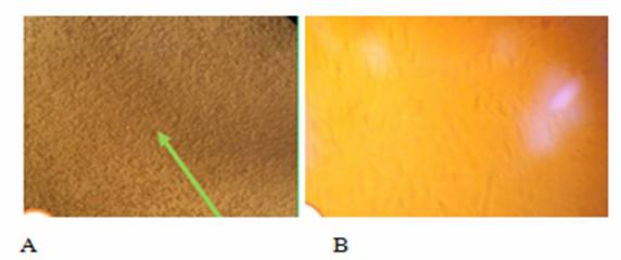
culture virus isolation showed that about 60% of inoculated BHK21 cells showed cythopathic effect and form rounding shapes. The previous
studies reported by different authors also indicated that FMDV cytopathic effect was characterized by inducing fast
destruction of BHK-21 cell monolayer. This was rationalized by (Shawky et al.,
2013) who stated virus isolated from clinical cases resulted in distortion of
the cell and cytopathic effect within 24-48 hours
after infection. The other clinical samples that didn't show cytopathic effect may be due to death of the virus during
transportation and processing.
The
subtyping analysis stated that about 40% of clinical samples contained the
virus and O serotypes were identified. This was substantiated with the report
of Ayelet, (2009) who identified serotype O (73.3%),
serotype A (19.5%), SAT-2 (4.1%), SAT-1 (1.8%) and C (1.4%) in the field
outbreaks. The studies conducted by (Tesfaye, 2014)
also indicated that serotype O (65.63%) was the dominant strain that circulated
in Addis Ababa, Debre Berhan
and Bishoftu areas. In line with this, studies
conducted by Noureldin and Elfadil
(2014) in Khartoum of Sudan indicated that serotype O (82.6%) and SAT-2 (40%)
were the main circulating FMDVs in cattle.
The current O serotype was identified from clinical samples such as
epithelium, probang and oral swabs at Welmera district.
This study was in line with the reports of Knowles (2010) who identified
serotype O from clinical samples during FMD field outbreaks. There is a
possibility of having more FMDV serotypes circulating in the area that could
not be detected during the study period. The viral genetic material was
detected in the clinical samples collected from field outbreaks. It was
appreciated that there was higher concentration of viral load in the epithelial
tissues and oro-pharyngeal fluids. This could be
justified by the evidence that epithelial tissues were taken from clinical
cases while probang samples were taken from
symptomatic and asymptomatic cases. The preferred site for virus detection and
multiplication is epithelial tissues and the oral mucosa for producing vesicles
(OIE, 2012). This indicated that there
was high load of viral RNA in the epithelial tissues than in the probang samples. Moreover, clinical samples that didn't
induce the pathologic effect could have been due to virus degradation during
transfer from the field and difficulties in maintaining the cold chain during
transportation (Mwiine et al., 2009)
CONCLUSION AND RECOMMENDATIONS
Foot and
mouth disease is a rampant problem in the current study area. This was further
confirmed through clinical investigation, serological screening, virological detection and molecular based diagnosis. Hence, the results of the current study
indicated about 38.6% of infected cattle showed clinical signs during FMD
outbreak. As a result of this, young calves mostly suffered from this disease in
the farm. The dominant serotype circulating in the area and responsible for the
recent outbreak was FMDV serotype O. In
conclusion, the presence of enormous susceptible animals, age variability,
breed susceptibility, poor prophylactic immunization, uncontrolled cross border
movement, and high contact of animals at common points were identified as major
risk factors that could resulted in the occurrence of foot and mouth disease.
Therefore, field outbreak investigation will be required to detect the
emergence of new strains in circulation and identification of the potential
risk factors as well as vaccine matching test should be conducted to apply
potent vaccines against the serotypes per areas.
Amarel, M., Owen, N.,
Ferris, N., Kitching, R. and Doel,
R. (1993): Detection of foot-and-mouth disease viral sequences in clinical
specimens and ethyleneimine-inactivated
Ayelet, G., Mahapatra, M., Gelaye, E., Egziabher, G., Rufeal, T. and Sahle, M. (2009): Genetic characterization of
foot-and-mouth disease viruses Ethiopia 1981-2007. J. Emerg Infect. Dis., 15(9):1409-1417.
Belachew,T. (2014):Seroprevalence and involvement of small ruminants in the
epidemiology of Foot and mouth disease and molecular characterization of
serotypes circulating in selected district of Gambela
region, MSc Thesis, Addis Ababa University, Faculty of Veterinary Medicine, Bishoftu, Ethiopia..
Bewket, S., Mesfin, S. and Gelagay, A.
(2012): Presentation on foot and mouth disease surveillance, laboratory and
diagnostic activities in Ethiopia. East African Region
Laboratory Network 3rd Annual Network Meeting, 5 March, Nairobi, Kenya.
Bhattacharya,
S., Pattnaik, B. and Venkataramanan,
R. (1996): Development and
application of sandwich enzyme-linked immunosorbent
assay (ELISA) for type identification of foot-and-mouth disease virus in direct
field materials.
Indian J. of Ani. Scie., 19:
66:1–9.
Callahan,
J. D., Brown, F., Osorio, F.A., Sur, J.H., Kramer, E. and Long, W. (2002): Use
of portable real time reverse transcriptase polymerase chain reaction assay for
rapid detection of FMD virus. J. Am. Vet. Med Assoc., 220 (11): 1636 42..
CSA. (2009):
Central Statically Authority, Federal Democratic Republic of Ethiopia, Central
Statically investigation, statically Abstract.
Rushton, J. (2002):
The economics of Foot and mouth disease. OIE scientific and Technical Rev., 3: 637-644.
Kafeero, H., Frank, M., Mwiine, N., Kalenzi, A. and Nanteza, A. (2016): Comparative detection of FMD virus by
reverse transcription loop mediated isothermal amplification assay and real
time polymerase chain reaction in Uganda, Int.
J. of. Biotech and
Food Science., 4: 22–33.
Kandeil,
A., ElShesheny, R., Kayali,
G., Moatasim, Y., Bagato,
O. and Darwish, M. (2013): Characterization
of the recent outbreak of FMD virus serotype SAT2 in Egypt.
Arch Virol., 158 (3):619–27.
Knowles, N. J., and
Samuel, A. R. (2003): Molecular epidemiology of foot-and mouth disease virus. Virus Res., 91:65–80.
Knowles, N.,
(2010): FAO World Reference Laboratory for foot and mouth disease, Molecular
epidemiology reports, viewed on 22 June 2011.
McLaws, M., Ribble, C., Martin, W. and Stephen, C. (2006): Factors
associated with the clinical diagnosis of foot and mouth disease during 2001
epidemic in UK. Preventive Veterinary Medicine.
77:65-81.
Mwiin,
N., Ayebazibwe, C.,Olaho Mukani, W., Alexandersen, S. and Balinda, N.
(2009): Serotype specificity of antibodies against FMD virus in cattle in
selected districts in Uganda. Transbound. Emerg.
Dis., 57: 365–374.
Noureldin,
M. and Elfadil, A. (2014): Prevalence and Risk
Factors of Foot and Mouth Disease of Cattle in Khartoum State Sudan. Master Thesis, Sudan University of Science and Technology.
OIE. (2004):
Foot and mouth disease. In: Manual of Standards for Diagnostic Tests and
Vaccines. 5th ed,
Office international des Epizooties (OIE), Paris, France. Pp:
.111-128.
OIE.(2012): Foot and
Mouth disease, Manual of Diagnostic Tests and Vaccines for Terrestrial Animals,
Version adopted by the World Assembly of Delegates of the OIE in May 2012, OIE Terrestrial. Pp. 1-29.
Paton D.J. (2005):
Selection of Foot and mouth disease vaccine strains :
a review .Rev Scie Tech 24,981-993
Quinn, J.,
Markey, K., Carter, E., Donnelly, C. and Leonard C. (2005): Veterinary
microbiology and microbial disease. Blackwell Science Ltd, A Blackwell
publishing company. Pp. 402-407.
Shaw, A., reid, E.,Ebert,
S.M., Hutchings, k., Ferris, G.H. and king,D.P.
(2007): Implementation of one step real time RT-PCR protocol for diagnosis of FMD .J.of.Virol.Meth., 143: 81–85.
Shawky, M., Abd .A. Fakry, M., Daoud, H.M., Ehab E.S., Wael, M. G., Rizk, S.A., Mohamed,A. and Farouk, E. M. (2013): Isolation and
Molecular Characterization of Foot and Mouth Disease SAT2 Virus during Outbreak
2012 in Egypt. J. Vet. Adv., 3(2): 60-68.
Tesfaye, Y.
(2014): Isolation, Molecular Characterization and Vaccine Matching of Foot and
Mouth Disease Virus Circulating in Ethiopia. MSc Thesis,
Addis Ababa University, Faculty of Veterinary Medicine, Bishofu,
Ethiopia.
Yang, M., Goolia, M., W, Bittner, H. and Clavijo
A. (2013): Development of a quick and simple detection methodology for FMD
virus serotypes O, A and Asia 1 using a generic rapid assay devie.
Virol. J., 10:125.