INTRODUCTION
African
Mahogany (Khaya grandifiolia): Local Names English (Nigerian
mahogany, ivory Coast mahogany, Gold Coast mahogany, African mahogany); French
(acajoud’ Affique, acajou); German (rotes-Khaya, mahagoni); Indonesian (kaya);
Trade name African mahogany.
(Opuni-Frimpong, 2006, Lemmens, 2008).
Khaya grandifiolia is distributed from Côte d’lvoire east to Cameroon and
south to Cabinda (Angola); it possibly also occurs in Guinea, Liberia, the
Central African Republic and Congo. It is fairly widely grown in plantations
within its natural area of distribution but also in tropical Asia and tropical
America.(Abdelgaleil et al., 2005)
Evergreen or deciduous or monoecious large to very large tree up to 60 m tall,
bole branchless for up to 30 m, usually straight and cylindrical, up to 160-210
cm in diameter, with large buttresses up to 2-4m high. Sometimes extending into
prominent surface roots; bark surface brown and slightly rough, exfoliating in
small circular scales leaving a pock-marked, mottled greyish brown and orange
brown surface, inner bark pink to reddish; crown massive and rounded twigs
glabrous. (Andre, 2011; Lemmens, 2008).
Leaves
arranged spirally but clustered near ends of branches, paripinnately compound
with 3-7 pairs of leaflets; stipules absent; petiole 1-4cm long, rachis 6-20cm
long: petiolules 0.5-1cm long; leaflets opposite, oblong to oblong-elliptical,
5-14cm x 2-6cm, cuneate to obtuse and slightly asymmetrical at base, distinctly
acuminate at apex, margins entire, leathery, glabrous, pinnately veined with
5-10 pairs of lateral veins inflorescence an axillary panicle up to 20cm long.
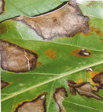
However,
since 1999 a high incidence of leaf spot caused by the fungus; Thanatephorus cucumers (teleomorph of Rhizoctonia
solani) has been observed, causing
numerous lesions on leaves of larger trees and 100% leaf fall in seedlings.
Seeds are commonly attacked by seed-boring beetles and eaten by small rodents.
Attacks of living trees by wood borers (Apate
spp.) have been observed. The bark of saplings is sometimes eaten by porcupines
and squirrels, which can kill the plants. In nurseries in Cote d’lvoire
seedlings are frequently attacked by psyllids (Phacosema spp.), bugs and scale insects, after which they are
infested by secondary fungal pathogens, resulting in a smut blackening the
leaves. (Abdelgaleil et al., 2005 and
Nwoboshi, 1982).
Fungal gummosis was initially observed in the
United States near Fort Valley, Georgia, during the 1960s. It subsequently spread
to other production areas of the Southeast. The disease was independently
discovered at about the same time in Japan, where it is described as peach
blister canker or ibokawa byo, and was later reported in China and
Australia. This disease, characterized by symptoms associated with bark
lenticels, contributes to a general decline of trees. (Weaver, 1974; 1979;
Beckman et al., 2003). Symptoms of fungal
gummosis are caused by a physiological race of Botryosphaeria dothidea (Moug.
Fr.). Other Botryosphaeria species have been reported to cause peach
gummosis in the United States (Pusey and Bertrand, 1993). However, these species are primarily wound
invaders that are not known to cause infections at lenticels. B. rhodina (Cooke)
Arx is relatively rare on peach. B. obtusa (Schwein.). Shoemaker
is very common in the Southeastern United States, but its importance as a peach
pathogen is unclear. B. obtusa is localized in dead tissue and outer
bark (rhytidome) and is absent or in the newly infected cortex and phloem
(Pusey et al., 1995; Weaver, 1974).
The earliest fungal gummosis symptoms appear
on young bark of vigorous trees as blisters 1 to 6 mm in diameter, generally
each with a lenticel at its center. These raised areas are due to an abnormal
multiplication of plant cells (Hyperplasia) in response to the causal organism
at the lenticel. Removal of outer bark
with a knife reveals diseased tissue at the lenticel margin surrounded by the
hyperplastic tissue. Blisters may be observed late in the season when infection
occurs or the following spring. By the end of the second season, the area of
necrosis surrounding lenticels has enlarged, and the hyperplasia is often less
visible or absent. Some second season necrotic lesions exude resins. Lesions
that appear in the second season after infection may or may not be preceded by
the formation of blisters (Pusey 1993; Pusey and Bertrand, 1993).
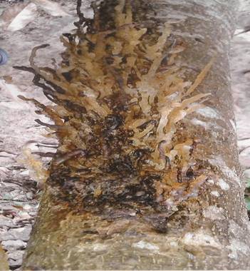
Beginning when trees are 2 or 3 years old,
sunken necrotic lesions encircling lenticels can be seen on the trunk and major
branches. Typically, copious resin exudate is associated with lesions at
multiple sites. Lesions of 2 cm or more in diameter on the oldest bark may
coalesce to form large cankers (Figure 2.1). Phloem and cortex are primarily
affected however, necrosis may extend to the xylem. Peach branch with blisters
caused by the fungus Botryosphaeria dothidea (Pusey et al., 1986)
The fungus overwinters in diseased bark and
in dead and dying wood, where it produces an abundance of spores. It spreads
within the orchard mainly by dispersal of conidia in rainwater. In the
Southeastern United States, asexual spores of the fungus are present from March
through October. Infections at lenticels develop from March through August, but
May through July is the key infection period. The fungus also invades through
wounds, causing cankers. Cankers may remain active for more than one year and
lead to secondary infections at lenticels. Blossoms, leaves, and fruits are not
infected (Weaver, 1974).
Regrettable
there are limited information about the diversity of Lasiodiplodia theobromae. Lack of information on host range of L. theobromae on the trees found in
Aboretum of Forestry and Environment, Rivers State University, Nkpolu-Oroworukwo,
Port Harcourt. Therefore, the present study was undertaken to observe the
influence of environmental factors on the mycelial growth of Lasiodiplodia theobromae.
This
research is aimed at investigating the pathological studies on Lasiodiplodia theobromae the causal
agent of gummosis infected African mahogany.
Specific
objectives of this research were to:
(i)
isolate and identify fungal pathogens
associated with infected leaves and stem bark of African mahogany.
(ii)
determine effect of temperature on the
mycelial growth of Lasiodipodia
theobromae.
(iii)
assess the effect of light and darkness on
mycelial growth of Lasiodiplodia theobromae.
MATERIALS AND METHODS
Study Area
The study was carried
out at the laboratory of Forestry and Environment (Pathology Unit) and Food
Science and Technology, Rivers State University, Nkpolu Oroworukwo, Port
Harcourt.
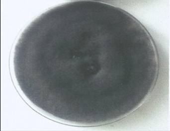
Sample Collection
The infected diseased
plant parts showing typical symptoms of gummosis disease were collected from
stem bark and leaves portions of diseased African mahogany tree from Aboretum
of Forestry and Environment, Rivers State University, Port Harcourt.
Effect of different temperature in the
mycelial growth of Lasiodiplodia
theobromae
The results on the
effect of different temperatures on Lasiodiplodia
theobrammae mycelial growth are presented in Table 2. The result showed
that different temperature and culture media influenced the mycelial growth of L. theobrammae. The relative increase in
fungus mycelial growth increased with the increase in temperature. It was observed
that the temperature range of 25-35oC was optimum for mycelial
growth in the PDA medium (15.6 ± 0.02mm
– 30.6 ±
0.05mm; 18.4 ± 0.28mm
– 32.5 ± 0.10mm).
Table 2: Effect of Different Temperature on
the mycelial Growth of Lasiodiplodia
theobromae (Mean ± SD)
|
Temperature
(toC)
|
Incubation period/mycelial growth (mm)/days
|
|
|
|
|
PDA
5 10
|
|
|
|
|
20
|
12.0 ± 0.01
|
14.6 ± 0.81
|
|
25
|
15.6 ± 0.02
|
18.4 ± 0.28
|
|
30
|
20.5 ± 0.03
|
24.0 ± 0.22
|
|
35
|
30.6 ± 0.05
|
32.5 ± 0.10
|
|
40
|
21.5 ± 0.06
|
23.5 ± 0.22
|
Mean
± SD (n=5) * PDA = Potato dextrose agar
Effect
of light and darkness on mycelial growth of Lasiodiplodia
theobromae on potato dextrose agar (PDA) incubated at room temperature (28 ± 2oC)
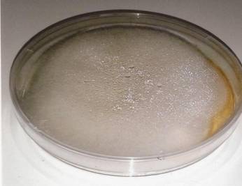
The result on the
effect of light and darkness in Lasiodiplodia
theobromae growth on stem bark tissues and leaves portions of Khaya ivorensis are shown in Table 3.
The result indicated that light and darkness significantly (p≤ 0.05)
affected the growth of L. theobromae at
different days. On the 5th day of incubation, L. theobromae under continuous darkness mycelial growth on PDA was
(13.5 ± 0.20mm – 18.2 ± 0.30mm). In continuous light, L. theobramae mycelial growth was (12.3 ± 0.02mm – 16.0 ± 07mm). Generally, the highest growth was observed after 10 days for
light and darkness on both PDA medium.
Table 3: Effect of light and darkness on
mycelial Growth of Lasiodiplodia
theobromae on PDA incubated at room temperature 28 ± 2oC (Mean ± SD)
|
Light and darkness
|
Incubation
period/mycelial growth (mm)/days
|
|
PDA
5 10
|
|
Continuous light
|
12.3 ± 0.02
|
16.8 ± 0.22
|
|
Continuous darkness
|
13.5 ± 0.20
|
18 ± 0.30
|
* PDA = Potato dextrose agar
DISCUSSION
The
results on the frequency of occurrence of fungal pathogens of infected stem
bark tissues and leaves of African mahogany (Table 1) indicated that Lasiodiplodia theobromae and Fusarium
solani were found to be responsible for the stem and leaves rot of African
mahogany collected from the forest arboretum. The stem and leaves pathogens of African mahogany have earlier been
implicated by some researchers. Ukoima and Chukunda (2016) reported on
the influence physiological factors on mycelia growth of B. thobromae, and Rhizopus
stolonifer caused serious rotting in Annoan muricata. Similarly, Ukoima et al., (2013) isolated Aspergillus
niger from seeds of Jatropha. This is in conformity with present research
work.
Chukunda (2014) found Aspergillus
niger, Aspergillus flavus, Rhizopus stolonifer, Fusarium pallidoroseum,
Botryodiplodia theobromae, Colletotrichum gloeosporoides, Penicillum expansum and
Botrytis cinera to be responsible for
the serious decay of avocado pears obtained in the Niger Delta ecosystem.
The
mycelia growth of lasiodiplodia
theobromae (Table 2) showed a variable trend in response to temperature
change using potato dextrose agar (PDA). Mycelia growth increased as
temperature increased from 20-35OC and then decreased with further
increase temperature. However, optimum mycelia growth of test fungus occurred
at 25-35OC. This results agreed with those reported by Quroshi and
Meah (1991) and Alam et al., (2001)
who reported that 25-30OC temperatures was optimum for the colony
growth and sporulation of lasiodiplodia
theobromae.
The
results of the effects of light and darkness on fungal growth (Table 3) revealed
that there was an increase in growth of L.
theobromae in both light and darkness. Rewal and Grewal (1989) studied the
effect of light on conidial germination of three strains of Botrytis cinerea infecting chickpea, and
found that conidia of B. theobromae germinated best under continuous
light and strain B2, B. theobromae of germinated well under
light and darkness treatment. From the study, it implied that light and
darkness are necessary for growth and sporulation of test fungi. This is in agreement with Ahmed (1985) who
observed that light promoted the growth and sporulation of Collectotrichum gloeosporoides.
Similarly, Marshi et al., (1959) reported that fungi exhibited varying response to
light depending on the light intensity, quality and duration of exposure. Prota (1992), Oladiran and Iwu (1993) and
Pihet et al., (2009) reported that
ultra violet (UV) radiation or sunlight affected the survival of fungal spores,
sclerotia and pycndia. However, some fungi need light to sporulate whereas
other fungi sporulate better in darkness.
In their investigation, Aspergillus
ornatus produced abundant conidia when grown in continuous light and
virtually none when grown in dark while Cleistothecia and ascospores are
produced in the dark whereas neither is produced in continuous light
(Schwemmin, 1960).
Hill
(1976) further explained that light inhibits glucose uptake and phosphorylation
which caused starvation and retards fungi growth and conidia formation.
Conversely the growth of Mycospharella
pinodes, Aspergillus niger increased when exposed to darkness. (Ukioma and
Chukunda, 2016).
On
the contrary, Alam et al., (2001)
reported that light is not necessary for growth and sporulation of B. theobromae,
but it enhances the growth and the number of conidia formation which is in
partial agreement with the observation of Rewal and Grewal (1989). The increasing
glucose in medium, the fungus could utilize it in a certain level and grow
properly, and after that level, the fungal physiology does not permit the
utilization of glucose for growth of the pathogen. There the fungus might utilize the glucose by
different ways instead of growth and formed more pigmentation using more
glucose.
According
to Cochrane (1958), temperature range permitting reproduction is usually
narrower than that permitting growth.
Earlier, Leach (1979) had reported variations in optimum temperature
requirements within the same species for light induced sporulation at
continuous light and continuous darkness Alam et al., (2001) obtained more growth of L. theobromae under continuous light and less in continuous darkness.
These findings agreed with the present research work where L. theobromae test fungi had a good growth performance for both
light and darkness.
However,
Teyegaga and Clerk (1972) earlier demonstrated the relationship between Cercospora canescens conidia longevity and storage humidity, and observed that
in the dark there was longest survival of conidia at low humidity than those
under light. Generally the spores
stored in the darkness appeared to be more viable than those in light. This may be due to metabolic disruption by
light or that light inhibited the spores of test fungi thus reducing their
percentage conidial germination.
CONCLUSION AND
RECOMMENDATIONS
Conclusion
Results from the
study revealed that Lasiodiplodia
theobromae is a major serious disease affecting the production of Khaya granifiolia from seedling level.
Temperature and light/darkness also played an important role in L. theobromae growth particularly
influenced by the culture medium growth kinetic. Results showed that L. theobromae was highest at a
temperature of 30-35OC.
Recommendations
Based on the present
findings the following recommendations are made;
1.
To reduce yield losses a good establishment
of host range of trees that are not susceptible to this pathogen should be
planted.
2.
Silvicultural practices will help reduce
re-occurrence of Lasiodiplodia theobromae
infection on the plantation.
3.
Preventing wounds on the test plant is the
best way to minimize the spread of L.
theobromae.
4.
Cankered branches of Khaya ivorensis should be pruned to reduce the inoculum from
initiating new infection.
5.
It is revealed from the study that
temperature, light and darkness significantly (P≤0.05) affected the
mycelial growth of L. theobromae.
RFERENCES
Abdelgaleil,
A. A. M Hashinaga, F & Nakatani, M (2005). Antifungal activity of limonoids
from Khaya. ivorensis. Pest Management Science, 61 (2):
186-190.
Alam,
S.M., Most-Ferdousi, B., Montaz, A.S., Rafiqul Islam, M. & Shah-Alam-M
(2001). Effect of Temperature, Light and Media on growth, sporulation,
formation of pigments and Pycnidia of Botryodiplodia
theobromae Pat. Pakistan Journal of
Biological Science, 4 (10):
1224-1227.
Andre,
R. E (2001). The development of community
forests in Cameroon: Origin, current situation and constraints. Networks
paper 25b.
Beckman,
T. G., Pusey, P. L. & Bertrand P. F. (2003). Impact of fungal gummosis on
peach trees. Horticultural Sciences,
38 (6): 1141-1143.
Chukunda, F. A
(2014). Post-harvest fungal diseases of Persea
gratissima (C.F. Gaerth) and Dacryodes
edulis (G.Don) H.J. Lam, Fruits. Rivers State, University of Science and
Technology, Ph.D Rivers State, Nigeria, 21.
Chukunda, F.A &
Offor, U. S (2015). Nutritional composition and fungal spoilage of African pear
(Dacryodes edulis) fruits sold
in Port Harcourt Metropolis Nigeria. Journal of Research in Biology, 5 (6)
1809-1814.
Cochrane, V. W (1958). Physiology
of fungal. John Wiley & Sons, Inc. New York, 524.
Lemmens,
R.H.M.J (2008). Khaya ivornisis A. Chev. In: Louppe, D. Oteng-Amoako, A.A.
& Brink, M. (Editors). PROTA (Plant Resources of Tropical Africa I
Resources vegetales del’Afrique tropicale, Wageningen, Netherlands. Accessed 8
September 2016.
Maheswari, S. K.,
Singh D. V. & Sahu A.K. (1999).
Effect of several nutrient media, pH and carbon sources on growth and
sporulation of Altemaria altemata. Journal of Mycopathology Research, 37,
21-23.
Oladiran, A. O. & Iwu, L. N
(1993). Studies on fungi associated with tomato fruit rots and effect of
environmental factors on storage. Mycologia,
21:157-163.
Opuni-Frimpong,
E (2006). Improving productivity and conservation of African mahogany: genetic
selection, propagation and silvicultural management of Hypsipyla robusta (Moore). PhD Forest Science
degree thesis, School of Forest resources and Environmental Science, Michigan
Technological University, Houghton, United States, 177.
Pathak,
V. N (1987). Laboratory manual of plant
pathology (2nd edition). Oxford and IBH publishing Co. Pvt. Ltd
New Delhi, India.
Punithalingam,
E (1976). Botryodiplodia theobromae, CMI
descriptions of pathogenic Fungi and Bacteri No. 519. Kew, Survey, UK, Common
Wealth Mycological Institute.
Pusey, P. L. & Bertrand, P. F. (1993). Seasonal
infection of nonwounded peach bark by Botryosphaeria dothidea. Phytopathology, 83: 825-829.
Pusey, P. L. (1993). Role of Botryosphaeria
species in peach tree gummosis on the basis of differential isolation from
outer and inner bark. Plant Disease,
77: 170-174.
Pusey, P. L., Kitajima, H & Wu, Y (1995). Peach tree fungal gummosis.
In: Compendium of stone fruit diseases. J. M. Ogawa, E. I. Zehr, G. W. Bird, D.
F. Ritchie, K. Uriu and J. K. Uyemoto, eds. APS Press, St. Paul, MN. 33-34.
Pusey, P. L., Reilly, C. C. & Okie, W. R. (1986). Symptomatic
response of peach trees to various isolates of Botryosphaeria dothidea. Plant Disease 70: 568-572.
Rewal, N. & Grewel, J. S
(1989). Effect of temperature, light and relative humidity on conidial
germination of three stains of Botrytis
cinerea infecting chickpea Indian
Phytopathology 42: 79-83.
Saleem A & Nasir, M. A.
(1991). Culture Media Directorate of Agricultural Information, Agriculture
Department, Government of the Punjab, Lahore.
Schwemmin, D. J
(1960). Light controlled
reproductive differentiation in
Aspergillus ornatus. University
of Michigan, U.S.A.
Steel,
R. G. D & Torrie J. H. (1980). Principles
and Procedures of Statistics: A Biometrical Approach. 2nd
edition. 597. McGraw Hill Book Company, New York.
Teyegaga, A. & Clerk, G. C (1972). Germination and survival of
conidia of Cercospora caneslens. Tropical Agriculture, 6: 197-204.
Ukoima
H. N. and Chukunda, F. A (2016). Influence of physiological factors on mycelial
growth of Botryodiplodin theobromae
Pat., isolated from Annona muricata. Nigerian Journal of Oil and Gas Technology, 1 (1): 39-50.
Ukoima,
H. N., Chukunda, F. A. & Etim, G (2013). Fungal seed borne disease of Jatropha
curcas and their in vitro control measures.
International journal of
phytofuels and other Allied Sciences, 1:29-35.
Weaver, D. J (1974). A
gummosis disease of peach trees caused by Botryosphaeria dothidea. Phytopathology 64: 1429- 1432.
: