|
Greener Journal of Medical Sciences
Vol. 11(1), pp. 46-72, 2021
ISSN: 2276-7797
Copyright ©2021, the copyright of this article is retained by the
author(s)
https://gjournals.org/GJMS
|

|
Prevalence and
Pattern of Dermatophytosis in Patients with Human Immunodeficiency Virus
Infection Seen in The University of Port Harcourt Teaching Hospital, (UPTH)
Port- Harcourt
Amaewhule MN
Department of Internal Medicine,
Rivers State University Teaching Hospital.
|
ARTICLE INFO
|
ABSTRACT
|
|
Article No.: 040421032
Type: Research
|
Background: Immunosuppression
due to various aetiologies have been associated with
the occurrence of dermatophytosis. Several studies in the past have
demonstrated that Human Immunodeficiency Virus (HIV) infection is a risk
factor for the acquisition and severity of dermatophytosis.
Aim: This study examined the prevalence and pattern of
dermatophytosis among patients with HIV infection in the University of Port -
Harcourt Teaching Hospital, Port Harcourt, Southern
Nigeria.
Method: This was a cross-sectional study carried
out in Port Harcourt over a 6 month period involving 173 HIV sero-positive
cases and 173 seronegative controls subjects. They were interviewed with a
structured questionnaire and thereafter screened for the presence of
dermatophytosis. Samples were collected from those with clinically diagnosed
dermatophytosis and sent for mycology studies. Information were
anaylsed using SPSS version 20.
Results: There was a higher prevalence of
dermatophytosis in the HIV seropositive group when compared to the HIV
seronegative control made up of HIV seronegative subjects. Most of the
lesions seen were not markedly different from that seen in immunocompetent
persons. 41.65% of the cases were found among those with Cd4 cell counts below
200cells/µl. Tinea corporis was the commonest lesion seen (50%). Trichophyton
species was the commonest dermatophyte isolated, followed by Microsporum
species.
Conclusion: The prevalence of dermatophytosis is
significantly higher in HIV infected patients and commonly occurs in advanced
stages of the disease. Tinea corporis is the most common lesion in this group
of patients and Trichophyton species a common causative agent.
|
|
Accepted: 05/04/2021
Published: 08/04/2021
|
|
*Corresponding Author
Amaewhule MN MBBS, FWACP
E-mail: nnendamary@ gmail.com
|
|
Keywords: prevalence;
pattern; dermatophytosis; HIV; UPTH
|
|
|
|
INTRODUCTION
Dermatophytosis
is a superficial fungal infection of the skin, hair or nails. Dermatophytes are
characterised by their ability to exist and grow in keratin, enabling them to
invade the stratum corneum of the skin and keratinized structures such as hair
and nails with minimal stimulation of the host’s immune response.
Dermatophytes belong to 3 genera:
-
Trichophyton
-
Microsporum
-
Epidermophyton
The
growth of dermatophytes in keratin is restricted to production of hyphae, which
branch and segment into chains of spores called arthrospores or arthroconidia.
Arthrospores are the main means of dissemination and propagation of the fungus
and can remain viable and in the environment and exfoliated skin for many
months and even years.
The type and extent of fungal invasion in
dermatophytosis of the hair as well as its clinical features differs according
to the species of fungi. The hyphae and arthrospores of some species remain
within the hair shaft (endothrix) while others form a sheath of arthrospores
around the shaft of the hair (ectothrix).
Dermatophytes grow best in a warm, humid
environment and are therefore more common in the tropical and subtropical
regions. The geographic distribution varies with the organism Microsporum canis, Microsporum nanum, Trichophyton
mentagrophytes, Trichophyton verrucosum and Trichophyton equinum occur worldwide1.
Trichophyton simii (found in monkeys) occurs only in Asia, and Trichophyton
mentagrophytes var. erinacei is limited to France, Great Britain,
Italy and New Zealand1.
Trichophyton schoenleinii and Trichophyton soudanense are
commonly found in Africa. In the West African sub-region, Trichophyton soudanense, Microsporum audounii, Microsporum canis, Trichophyton
violaceum, and Trichophyton rubrum
are common aetiological agents of dermatophytosis1,2.
There are about 40
recognised specie of dermatophytes. Some are only able to infect man
(anthropophilic), others are primarily animal
pathogens (zoophilic) but can also infect man. Other species are found as
saprophytes in the soil (geophilic), and cause sporadic infection in man and
animals.
Dermatophytosis are mild communicable diseases with high
morbidity and contribute to major health problems in the tropics and sub
tropics especially in Nigeria. Most surveys on dermatophytosis done in the past
have been carried out in school children and have concentrated mainly on tinea
capitis. This maybe because of the social stigma attached to it, possibility of
alopecia and other associated secondary diseases such as bacterial infection of
the lesions.
The prevalence rate of dermatophytosis in the
general population in a study done in Lagos is about 6.1%3.
Transmission: Infection occurs by contact with
arthrospores (asexual spores formed in the hyphae of the parasitic stage) or
conidia (sexual or asexual spores formed in the “free living” environmental
stage). Infection usually begins in a growing hair or the stratum corneum of
the skin. Dermatophytes do not generally invade resting hairs, since the
essential nutrients they need for growth are absent or limited. Hyphae spread
in the hairs and keratinized skin, eventually developing infectious
arthrospores. Transmission between hosts usually
occurs by direct contact with a symptomatic or
asymptomatic host, or direct or airborne
contact with its hairs or skin scales. Infective spores in hair and dermal scales can remain viable for several months to years in the environment4.
Geophilic dermatophytes, such as Microsporum
nanum and Microsporum gypseum, are usually acquired directly from
the soil rather than from another host4.
Fomites are also another important means of transmission5,6.
Factors affecting
infection:
Factors
inhibiting the growth of dermatophytes include saturated fatty acids in
sebaceous glands7.
Neutrophils
and monocytes have been found to kill dermatophyte conidia, a process which
depends on both intra-and extracellular mechanisms8.
Antibodies to dermatophytes have not been
found to be protective9; however development of cellular immunity
via sensitized T-lymphocytes is a key factor in immunological defence10.
Host immunity against dermatophytosis depends on both innate and acquired
immune mechanism. Chronic infections are associated with poor T-lymphocyte
function11,12.
Other host factors affecting infection are as
follows- genetic susceptibility, reduced immunity in old age, diabetes
mellitus, Cushing’s syndrome, and HIV infection13-17.
Environmental factors like humidity and
raised CO2 tension favour dermatophyte invasion18. Raised
temperature of more than 37oC inhibits dermatophyte growth19.
This is partly
responsible for the lack of deeper penetration of the skin in dermatophytosis.
Clinical
Features:
The
disease produced by dermatophytes are described according to anatomic site
involved viz – tinea capitis (scalp), tinea barbae (bearded skin of the face),
tinea corporis (the body), tinea cruris (groin), tinea unguium ( the nails),
tinea manuum (hand) and tinea pedis (the feet). These infections may vary from
mild inflammations to acute vesicular reactions.
The incubation period in humans is 1 to 2 weeks.
Tinea Capitis: This occurs
predominantly in prepubertal children. Adult infection is rare. One risk factor
for adult infection is immunosuppression from HIV or drugs. Microsporum and
Trichophyton species are the main aetiological agents. The most common
causative fungi is Trichophyton
tonsurans and Microsporum canis.
Lesions vary from a dry, scaly patch of alopecia to the development of pustules
and abscesses also known as kerion.
Tinea Barbae: This refers to
dermatophytic infection of the bearded area of the face. It may present as
folliculitis or as a severe inflammatory reaction consisting of papules,
pustules, exudates and crusting. Common causative organisms include Trichophyton veruccossum, Trichophyton
violaceum, Trichophyton mentagrophytes, Trichophyton schoenleinii, Microsporum
canis and Trichophyton rubrum.
Tinea Corporis: This refers to
dermatophytosis of the skin excluding the hair, nails and feet. Skin lesions
may be dry and scaly or moist and crusty. As they enlarge their centres heal
producing the classic annular lesions.
Lesions may also be pustular, vesicular and occasionally granulomatous.
All
dermatophytes can produce tinea corporis. Tinea incognito occurs if a topical
steroid has been applied and the clinical appearance of the initial tinea
lesion is altered, becoming less scaly, more extensive, pustular, pruritic, and
painful.
Tinea Cruris: Dermatophytosis of the groin area. Infections occurs in proximal thighs, crural folds and
extends onto the buttocks. Lesions are raised, sharply defined, erythematous
and pruritic.
Common
causative organisms include: Trichophyton
rubrum, Epidermophyton floccosum, Trichophyton mentagrophytes var
interdigitale.
Tinea Pedis: This refers to
dermatophytosis of the foot including the plantar surface and toe web space. It
presents with itching pain, maceration, and hyperkeratosis of soles and sides
of the feet.
Causative
organisms include Trichophyton rubrum, Trichophyton mentagrophytes
and Epidermophyton flocossum.
Tinea Manuum: Dermatophytosis of
the hand especially involving the palmar surface, it presents as a dry scaling
eruption, may also presents as ulcerations and
hyperkeratosis. Trichophyton rubrum
is commonly implicated.
Tinea Unguium: Dermatophytosis of
the nail plate. Also called onychomycosis. It usually
starts at the tip of one or more nails. There is a gradual thickening,
discoloration and crumbling of the affected nail which may eventually be
completely destroyed. Causative fungi include Trichophyton rubrum, Trichophyton mentagrophytes var interdigitale and Epidermophyton floccosum.
Chronic dermatophyte infections may be the
first manifestation of HIV and may suggest HIV infection because of increased
severity of presentation, atypical clinical appearance or increased resistance
to treatment.
The burden of HIV: HIV infection is a
major challenge and health problem worldwide. It was first discovered in Los
Angeles, California in young
homosexual men who presented with disseminated Kaposi sarcoma and pneumocystic
carinii infection in 198120,21,22.
It
later became evident that this disease affected other population groups as well
when some cases were reported in intravenous drug users. In 1983, almost 2
years later HIV
was defined as the primary cause of Acquired Immunodeficiency Syndrome (AIDS)23,24,25.
Since then it has become a global problem with rising incidence in various
parts of the world.
Worldwide more than 34 million people are
infected with 69% in Sub Saharan Africa. Globally, 0.8% of adults aged 15-49
years are living with the virus26.
The
prevalence rate for HIV in Nigeria is 4.1%27 previously, now 1.4%.26
The geo-political zone with the highest HIV
sero-prevalenceis the North-central zone (7.5%); while the North-western zone
has the lowest prevalence rate of 2.1%27. Benue state in the North
central zone has the highest prevalence of 12.7% while Kebbi state in the
North-western zone has the lowest prevalence of 1.0%27. A total of
3,459,363 are currently infected with the virus in this country27.
Urban areas have a higher prevalence than rural areas27. Women,
youths, and people with a low level of education are worst affected27.
Human
Immunodeficiency Viral infection are commonly associated with a myriad of skin conditions.
Examples include pruritic papular eruption, herpetic infections, Kaposi sarcoma
and dermatophytosis and these skin conditions may be the first manifestation of
the disease.
The development of cellular immunity via
sensitised T-lymphocytes is a key factor in immunological defence against
dermatophytic infections. HIV alters this defence by producing cellular immunodeficiency
characterised by the depletion of Helper T-lymphocytes (CD4 cells) thereby
predisposing to dermatophyte infections.
Animal models have shown that Langerhan’s
cells which act as antigen presenting cells to dermatophyte antigen are the
first cellular targets of HIV. The virus fuses with these cells and spreads
into deeper tissues28,29. The virus has
also been identified in epidermal Langerhan cells in HIV infected patients30,
and these patients have a reduced number of such cells which leads to a
compromise in the skin immune response which may result in multiple or
extensive skin infections.
Acquired immune
deficiency syndrome (AIDS) was first discovered as a novel disease in 1981.21
Within 2 years of defining AIDS as a distinctive syndrome in 1981, the human
immunodeficiency virus (HIV) was identified as the causative agent. HIV
infection is acquired sexually, from blood or blood products, or vertically
from an infected mother
to her child during pregnancy, delivery or breastfeeding. The
virus infects immunocompetent cells including CD4 T-cells and macrophages. It
creates variable patterns of disease in individuals, groups and races. These
diseases are characterized by evolving, sometimes fulminant immunodysfunction
(AIDS) affecting many
systems of the
body.
Aetiology:
HIV belongs to the lentivirus group of the retrovirus
family. There are two types, HIV-1 and HIV-2. HIV-1 is the most frequently
occurring strain globally. HIV-2 is almost entirely confined to West Africa,
although there is evidence of some spread to Europe, particularly France,
Portugal and the Indian subcontinent31.
HIV-2 has only 40% structural homology with
HIV-1 and although it is associated with immunosuppression and AIDS, appears to take a more indolent course than HIV-1. Many of the drugs that are used in HIV-1 are
ineffective in HIV-2. The structure of HIV is shown
in
figure 1 below.

Figure 1:
Diagram of Human Immunodeficiency Virus.
Retroviruses are characterized by the possession of
the enzyme reverse transcriptase, which allows viral Ribonucleic Acid (RNA) to
be transcribed into Deoxyribonucleic Acid (DNA), and thence incorporated into
the host cell genome. Reverse transcription is an error-prone process with a
significant rate of mis-incorporation of bases. This, combined with a high rate
of viral turnover, leads to considerable genetic variation and a diversity of
viral subtypes or clades. Upon entry into the target cell, the viral RNA genome is converted
(reverse transcribed) into double-stranded DNA by a virally encoded reverse
transcriptase
that is transported along with the viral genome in the virus particle. The
resulting viral DNA is then imported into the cell nucleus and integrated into
the cellular DNA by a virally encoded integrase and host co-factors.
Once integrated, the virus may become latent, allowing the virus
and its host cell to avoid detection by the immune system. Alternatively, the
virus may be transcribed, producing new RNA
genomes and viral proteins that are packaged and released from the cell as new
virus particles that begin the replication cycle anew. On the basis of DNA sequencing, HIV-1 is
divided into three groups (M, N, and O), which probably represent three
zoonotic transfers from the chimpanzee but do not differ clinically in humans32.
Group M (major) subtypes (95% of
infections worldwide) contains
at least 8 subtypes (or clades), which are denoted A–J33. There is a
predominance of subtype B in Europe, North America and Australia, but areas of
central and sub-Saharan Africa have multiple M subtypes. Subtype G is
predominant in Nigeria. Recombination of viral material generates an array of
circulating recombinant forms (CRFs), which increases the genetic diversity
that may be encountered.
Group N (new) is mostly confined to parts
of West-Central Africa (e.g. Gabon).
Group O (outlier) subtypes are highly
divergent from group M and are largely confined to small numbers centred in
Cameroon.
Transmission:
Despite the fact that HIV can be isolated from a wide
range of body fluids and tissues, more than 90% of HIV infections are transmitted via semen,
cervical secretions and blood.
Sexual intercourse (vaginal and anal):
Globally, heterosexual intercourse accounts for the
vast majority of infections, and coexistent sexually transmitted infections
(STIs), especially those causing genital ulceration, enhance transmission34,35. Transmission of HIV appears to be more
efficient from men to women, and to the receptive partner in anal intercourse,
than vice versa. In the United States, as of 2009, most sexual
transmission occurred in men
who had sex with men with this population accounting for 64% of all
new cases36. In central and sub-Saharan Africa (Nigeria inclusive) the epidemic has
always been heterosexual and more than half the infected adults in these
regions are women26. SouthEast Asia and the Indian subcontinent are
experiencing an explosive epidemic, driven by heterosexual intercourse and a high incidence of other sexually
transmitted diseases37.
Mother-to-child (transplacentally, perinatally, breastfeeding):
Mother-to-child transmission is the third most common
route of HIV infection globally34. Studies suggest that, without
intervention,
In the absence of treatment, the risk of transmission
before or during birth is around 20% and in those who also breastfeed 35%36. As of 2008, vertical transmission accounted for
about 90% of cases of HIV in children36. With appropriate treatment
the risk of mother-to-child infection can be reduced to about 1%36. In the developed world interventions to reduce
vertical transmission, including the use of antiretroviral agents, delivery by
caesarean section and the avoidance of breast-feeding have led to a dramatic fall in the
numbers of infected children. However, with the advent of highly active
anti-retroviral therapy (HAART), mothers are encouraged to exclusively
breastfeed their babies as the benefits outweigh the risks.
Contaminated
blood, blood products and organ donations: Screening of blood and blood products was introduced
in 1985 in Europe and North America. Prior to this, HIV infection was
associated with the use of blood products (in haemophiliacs) and with blood
transfusions. In some parts of the world where blood products may not be
screened, and in areas where the rate of new HIV infections is very high,
transfusion-associated infections continues to occur. HIV is
transmitted in About 93% of blood transfusions
involving infected blood39.
Contaminated needles (intravenous drug misuse,
injections, needle-stick injuries):
The practice of sharing needles and syringes for intravenous drug use continues
to be a major route of transmission of HIV in both developed countries and
parts of South East Asia, Latin America and the states of the former Soviet
Union. In some areas, including the UK, successful education and needle
exchange schemes have reduced the rate of transmission by this route.
Iatrogenic transmission from needles and syringes used in developing countries
is reported. Healthcare workers have a risk of approximately 0.3% following a
single needle-stick injury with known HIV-infected blood40.
Immunology And Pathogenesis Of HIV Infection: Primary HIV infection results in natural or innate
immune responses that are mobilized within hours of infection and include
inflammation, non-specific activation of macrophages, natural killer cells and
complement, and release of cytokines. After antigenic stimulation, acquired
immune responses are primed. These responses emerge at the same time as
clearance of viraemia and rebound of CD4 T cells is seen. These HIV-specific
responses include specific humoral or antibody responses and specific cellular (T-lymphocyte) responses.
Specific humoral or antibody responses41: This consist of neutralizing antibodies to the envelope
proteins of the virus and other non-neutralizing antibodies to internal viral
proteins such as gag. Specific secretory Immunoglobulin A (IgA) mucosal
antibodies are also produced. Neutralizing antibodies are usually measurable by
12 weeks after infection.
In specific cellular
(T-lymphocyte) responses42, CD8 T lymphocytes or cytotoxic-lymphocytes (CTLs) form
a primary component of the critical cellular immune response induced by HIV
infection. CTLs are differentiated from existing CTL precursors, and express
T-cell receptor molecules capable of recognizing specific viral epitopes
presented in the context of human leukocyte antigen (HLA) or Major
Histocompatibility Complex (MHC) molecules at the surface of infected target
cells. Mature CTLs are functional 5–10 days after antigenic stimulation,
recognizing, binding and then lysing the infected target cell. Virus-specific
CTLs evolve faster than antibody responses and are often induced before
seroconversion and before viral RNA has reached peak titres. Thus CD8 CTLs are
temporally associated with the fall in viraemia during acute infection, and
there is good evidence that CTLs play a major role in the control of HIV
infection at this time and later in HIV disease. Evidence for strong CD8
antiviral pressure can be appreciated by the number and variety of strategies
which viruses have evolved to avoid apoptosis and CTL recognition, thus
prolonging the life of the virally infected cell and enabling viral replication and dissemination43.
In addition to the lysis of infected cells, CD8 T cells can reduce viral replication
by the production of soluble factors. These factors are not antigen specific
but their production requires specific T-cell activation. Anti-HIV effects have
been found for interferon (IFN) interleukin (IL)-10, IL-13, IL-16 and the C-C
chemokines, macrophage
inhibitory protein-1β, (MIP-1β) and regulated upon activation, normal
T-cell expressed and secreted (RANTES). Such soluble factors may also have
profound effects on other opportunistic infections including those affecting
the skin. CD4 T-cell responses induced by HIV infection provide help to both
HIV-specific CTLs and B cells. CD4 T-helper cells recognize antigen in the
context of HLA class II molecules on the surface of antigen-presenting cells
such as dendritic cells. CD4 responses to a variety of HIV proteins (including env,
gag and nef) have been demonstrated in early disease, but
immunological abnormalities in T-helper function occur very early in HIV
infection, even before CD4 T-cell numbers diminish in the peripheral blood.
Furthermore, advances in the understanding of HIV-1 pathogenesis reveal that
mucosal tissues, primarily in the gastrointestinal tract, are major sites for
early viral replication and CD4 T-cell destruction, and this may represent the major viral reservoir44. Reduced proliferative capacity and diminished
IL-2 production in response to stimulation by exogenous antigens (including
those from HIV and other pathogens) is one of the hallmarks of HIV disease.
On recognition of their specific
antigen, naïve CD4 T cells differentiate from a common (Th0) precursor into
T-helper (Th)1cells, which differentially secrete
interleukin 2 (IL-2), IFN-γ, transforming growth factor-β and IL-12
and can activate macrophages and ‘help’ CTLs, or into Th2 cells, which secrete
IL-4, IL-5, IL-6 and IL-10 that can activate B cells to proliferate and
differentiate into antibody-producing plasma cells. Central to the cellular
immune response is the dendritic cell, which is the most potent
antigen-presenting cell. However, such cells on mucosal surfaces (Langerhans’
cells) may be some of the first targets in transmission; as well as
transporting viral antigens across mucosal barriers and presenting them to CD4
cells, dendritic cells may themselves become infected with HIV, and their
function compromised.

Figure 2: Schematic depiction of HIV attacking a CD4
lymphocyte
Clinical Features of HIV infection:
WHO Clinical Staging of HIV/AIDS and Case Definition:
Clinical staging and
case definition of HIV for resource-constrained settings were developed by the
World Health Organisation (WHO) in 1990 and revised in 2007. Staging is based
on clinical findings that guide the diagnosis, evaluation, and management of HIV/AIDS, and it does not require a CD4 cell count. This
staging system is used in many countries to determine eligibility for
antiretroviral therapy, particularly in settings in which CD4 testing is not
available. Clinical stages are categorized as 1 through 4, progressing from
primary HIV infection to advanced HIV/AIDS (see Table 1 below). These stages
are defined by specific clinical conditions or symptoms.
TABLE 1: WHO CLINICAL
STAGING OF HIV/AIDS FOR ADULTS AND ADOLESCENTS45
|
|
|
 Asymptomatic
Acute retroviral syndrome
|
|
Clinical Stage 1
|
|
Asymptomatic
Persistent generalized lymphadenopathy
|
|
Clinical Stage 2
|
|
Moderate unexplained weight loss (<10% of
presumed or measured body weight)
Recurrent respiratory infections (sinusitis, tonsillitis,
otitis media, and pharyngitis)
Herpes zoster
Angular cheilitis
Recurrent oral ulceration
Papular pruritic eruptions
Seborrheic dermatitis
Fungal nail infections
|
|
Clinical Stage 3
|
|
Unexplained severe weight loss (>10% of presumed
or measured body weight)
Unexplained chronic diarrhea for >1 month
Unexplained persistent fever for >1 month
(>37.6ºC, intermittent or constant)
Persistent oral candidiasis (thrush)
Oral hairy leukoplakia
Pulmonary tuberculosis (current)
Severe presumed bacterial infections (e.g.,
pneumonia, empyema, pyomyositis, bone or joint infection, meningitis,
bacteremia)
Acute necrotizing ulcerative stomatitis, gingivitis,
or periodontitis
Unexplained anemia (hemoglobin <8 g/dL)
Neutropenia (neutrophils <500 cells/µL)
Chronic thrombocytopenia (platelets <50,000
cells/µL)
|
|
Clinical Stage 4
|
|
HIV wasting syndrome, as defined by the CDC
Pneumocystis pneumonia
Recurrent severe bacterial pneumonia
Chronic herpes simplex infection (orolabial,
genital, or anorectal site for >1 month or visceral herpes at any site)
Esophageal candidiasis (or candidiasis of trachea,
bronchi, or lungs)
Extrapulmonary tuberculosis
Kaposi sarcoma
Cytomegalovirus infection (retinitis or infection of
other organs)
Central nervous system toxoplasmosis
HIV encephalopathy
Cryptococcosis, extrapulmonary (including
meningitis)
Disseminated nontuberculosis mycobacteria infection
Progressive multifocal leukoencephalopathy
Candida of the trachea, bronchi, or lungs
Chronic cryptosporidiosis (with diarrhea)
Chronic isosporiasis
Disseminated mycosis (e.g., histoplasmosis,
coccidioidomycosis, penicilliosis)
Recurrent non_typhoidal Salmonella bacteremia
Lymphoma (cerebral or B-cell non-Hodgkin)
Invasive cervical carcinoma
Atypical disseminated leishmaniasis
Symptomatic HIV-associated nephropathy
Symptomatic HIV-associated cardiomyopathy
Reactivation of American trypanosomiasis
(meningoencephalitis or myocarditis)
|
HIV infection
produces a panorama of mucocutaneous manifestations, from the macular roseola –
like rash seen with the acute sero-conversion syndrome to an array of severe
and extensive skin lesions seen as the disease progresses46.
Olumide et al, in a seroprevalence survey of
HIV I, HIV II and Human T-Lymphotrophic Virus-1(HTLV I) among patients with
skin disease and Sexually Transmitted Disease (STD) in a dermatology clinic
found a rising seroprevalence to HIV I and HIV II over a 2 year period. This
further highlights the increasing trend in HIV associated skin disease47.
In another study to access the changes in the pattern of skin
disease in Kaduna North Central Nigeria over a 6 year period. HIV
related skin disease constituted about 4.3% of all the cases of skin diseases
seen48. In a study by Ogunbiyi et al to assess the
prevalence of skin disease in Ibadan, Nigeria it was also revealed that there is an increase in HIV
associated skin disease49.
Fungal infections are a common complication
in HIV infection and include dermatophytosis, deep mycosis and yeast infections50.
Nnoruka et al in a study to access the
pattern of skin disease in HIV positive patients and their correlation with CD4
cell counts found out that dermatophytosis constituted 24.3% of all cases of
skin diseases seen in this group of patients. Four hundred and seventy-seven
HIV sero-positive patients were used in the study. The mean CD4 cell count of
patients with dermatophytosis was 437.3+177 cells/µl51.
In a
study in India by Shobhana et al52, 410,
HIV seropositive patients were screened for skin disease. It was found that 40%
had mucocutaneous involvement at presentation. Mean age of the study population
was 29 years and male to female ratio was 2.5:1. The common mucocutaneous
morbidities detected include oral candidiasis (36%), dermatophytosis and
gingivitis (13% each), herpes zoster (6%), herpes simplex and scabies (5%
each). A striking feature noted in 36% of the males was straightening of the
hairs. Genital herpes was the commonest genital ulcer disease. Lesions
associated with a declining immunity include oral candidiasis, oral hairy
leukoplakia and herpes zoster with median CD4 cell counts of 94, 62 and 192 cells/µl
respectively. It was concluded from his study that a recognition of the protean
mucocutaneous diseases in HIV/AIDS helps in earlier diagnosis of HIV and serve
as a measure of the immune status of the individual.
Dermatophytoses are common cutaneous fungal infections in HIV infected
patients and can occur at any stage of the illness, and show
clinical variations50,53.
In the immunocompetent host, various risk factors have been identified for the
acquisition of dermatophytosis. These include poverty54,55,
close contact with animals and soil55-57. (especially
for geophillic and zoophillic dermatophytes). Other risk factors include male
sex57, poor sanitary conditions58,55.
Use of poorly sterilised barbing equipment have also been identified as an
important risk factor for transmission of tinea capitis in this environment and
in one study was responsible for the high prevalence of tinea capitis in the
community59. An important risk factor for tinea pedis infection is
the frequent wearing of occlusive footwears. The warm humid environment
surrounding the feet provides a conducive environment
for the growth of dermatophytes for this group of persons60.
Invasion of the skin by dermatophytes begins
with adherence of arthroconidia to the keratinocytes, followed by penetration
through and between cells and development of a host response61.
Adherence of dermatophytes to keratinocytes
takes about 2 hours to complete, during which germination and penetration of
the keratinocytes occur. Hyphal prolongation follows shortly afterwards and
proceeds radially61,62. The limitation of
invasion of dermatophytes to the stratum corneum is due to the presence of a
fungistatic factor in the tissue fluids and serum63,64.
Dermatophytes produce a variety of
proteolytic enzymes which play a role in the invasion of the stratum corneum,
hair and nails65-68. There is some heterogeneity in substrate
preference of dermatophytes. While all dermatophytes invade stratum corneum,
different species vary widely in their ability to invade nail and hair.
Trichophyton
rubrum
rarely invades hair but frequently invades nail. Epidermophyton. floccosum
never invades hair and only occasionally invades nail69.
The commonest clinical presentations of
dermatophytosis in immunocompetent persons are tinea corporis and tinea capitis69.
Various studies have demonstrated unusual
clinical presentations and higher prevalence rates of dermatophytosis among HIV
seropositive patients. In one study by Goodman et al, it was 4 times higher in
HIV positive patients59. In that study, 117 HIV seropositive
patients were recruited. Dermatophytosis was seen in 30% of the patients. Other
common skin diseases seen were: candidiasis (47%), seborrheic dermatitis (32%),
acquired icthyosis or xerosis (30%) and herpes simplex infection (22%).
Kaviarasan et al70, studied the
prevalence and clinical variations of dermatophytosis in HIV infected persons . A total of 185 HIV infected persons were screened
and a diagnosis of dermatophytosis was made in 41 cases. Prevalence of
dermatophytosis was 22.2%. Male: female ratio 3:1, mean age of the patients was
30.7 years. Tinea Corporis was the commonest dermatophyte infection (53.7% of
cases) followed by tinea cruris (49.9%), tinea pedis (17.1%), tinea facei
(14.7%). Tinea manuun was noted in 0.5%
while 6% of the cases had tinea unguium.
Out of the 22 patients with tinea coporis 19
(8.36%) were staged as full blown AIDS - WHO Clinical Stage IV. Ten (45.45%) of
them presented with the anergic form of tinea corporis, proximal subungual
onychomycosis, thought to be pathognomonic of AIDS was seen in 3 cases.
In another study by Ekong et al (utilising 4
centres in Nigeria)71. to
evaluate the types and clinical presentations of superficial and deep fungal
infections in HIV/AIDS patients, 288 patients were screened, aged 37±14.5
years. Mean baseline CD4 cell count was 450 cells/µI.
It was found that 69% of the patients had dermatophyte infections 35% with
tinea corporis, 28% with tinea pedis. Patient with low CD4 count had more
severe fungal infections while those on Anti-retroviral (ARV) drugs had milder diseases.
Dermatophytosis in HIV infection can present
as atypical lesions due to immunodeficiency. Skin lesions may be disseminated,
facial tinea mimicking seborrheic dermatitis, palmoplantar lesions with
significant hyperkeratosis, lesions practically without erythema and with a
prevalence of desquamation simulating xerosis72,73.
In HIV dermatophytosis can also present as
extensive and deep lesions74. A case of an HIV positive 23 year old
male was reported with a CD4 cell count of 335 cells/µl with
multiple large erythematous circinate and pustular plaques on his
abdomen, back, arms, and legs. Trichophyton
mentagrophyte was isolated and biopsy showed suppurative deep
dermatophytosis and folliculitis. The patient responded to itraconazole therapy
for 2 weeks74.
In another report by Kwon et al75,
a 44 year old HIV positive man with Trichophyton
rubrum infection presenting with widespread invasive multiple tumor
like eruptions. The patient responded to oral terbinafine therapy with complete
remission of his lesions.
Raquel et al also presented a case of an HIV
positive 33 year old Brazilian woman with an exacerbated inflammatory response
to Trichophyton rubrum infection of her left arm. This patient was treated with
oral fluconazole which she responded to76.
Uncommon dermatophytes such as geophillic and
zoophillic organisms have also been detected as causative agents of tinea
corporis in HIV infected patients. In a study by Porro et al, tinea corporis infection with atypical presentation caused by Microsporum gypseum, a geophillic dermatophyte were reported
in 2 patients with AIDS. Microsporum gypseum, is an
unusual causative organism for human dermatophytosis77.
In another report by Nenoft et al, an HIV
positive 15 year old boy from Uganda was found to have several dry and
hyperkeratotic lesions of the forearms and left hand with circinate,
erythematous and scaly morphology. Microsporum
gypseum was also isolated in this patient as a causative organism of this
tinea coporis78.
Nunman
et al also reported another similar case of extensive Tinea corporis infection
caused by Microsporum gypseum in a 36
year old HIV positive woman. The lesions were said to be generalised,
psoriasiform and refractory to treatment with ketoconazole and itraconazole79.
Zoophilic organisms have also been reported
as causative agents of tinea corporis in HIV infected patients. Menon et al
reported a case of an HIV infected patient who presented with a chronic non
inflammatory non pustular extensive infection caused by the zoophillic
dermatophyte Trichophyton verrucossum80.
Lowinger Seoane et al also described another
case of disseminated cutaneous dematophytosis caused by another zoophillic
organism. Trichophyton mentagrophytes
and Microsporum canis in an HIV
infected patient81.
Tinea capitis is rare in adults. This may be
due to the fact that the quantity of fungistatic saturated fatty acids in sebum
increases in puberty82.
It has also been found that dermatophyte
colonisation of hair disappears at puberty, this may result from the
colonisation by pityrosporum orbiculare
interfering with dermatophyte contamination, and the thicker calibre of adult
hair may protect against dermatophyte invasion82,83.
Tinea capitis in adults generally occurs in
patients who are immuncompromised and those infected with HIV84. It
is uncommon in immunocompetent adults and when it occurs the clinical features
may be atypical and this may delay the diagnosis84. It
may resemble bacterial folliculitis, folliculitis decalvans, dissecting
cellulitis or the scarring related to discoid lupus erythematosus84.
However, tinea
capitis in men even if HIV positive is uncommon. A few cases of tinea
capitis have been described
in HIV infected
patients. Lateur et al presented 2 cases of adult black African males with HIV
infection with tinea capitis85.
In another report, by Bournerias et al86, 2 HIV sero-positive men presented with
unusual M.icrosporum canis infection.
Both had tinea capitis presenting as alopecia in one and scaling of the scalp
in the other. One also had tinea unguium caused by Microsporum canis. Both were treated with oral itraconazole for
several months and only one was cured.
One of the earliest superficial fungal
infections to emerge in HIV infected persons is onychomycosis. These group of patients are more likely to develop it when
the CD4 lymphocyte level falls to approximately 400 cells/µl87.
In the HIV positive
patient prevalence of onychomycosis ranges from 11% to 67%88. It often starts as a
proximal white subungual onychomycosis (PWSO) and quickly spreads to the other
nails of fingers and toes88,89,90. If left untreated it can lead to systemic
infection to which the immunocompromised host cannot respond.
Proximal subungual onychomycosis is said to
be an indicator of HIV disease91,92. Although it can also occur in other immunocompromised patients.
Another common presentation of onychomycosis
in HIV infected persons is the one hand, 2 feet tinea (that is affectation of
one hand and two feet) which is relatively uncommon in the general population91,92.
In a study of onychomycosis in 62 HIV
infected persons, Dompmatin et al found out that the most frequent aetiologic
agents were dermatophytes (in 58%). The rest are caused by candida albicans and Pityrosporum. Ovale93.
Proximal white subungual onychomycosis in HIV
patients is generally caused by Trichophyton
rubrum92,93. However, there are rare
reports of lesions caused by other species such as Trichophyton mentagrophytes, Epidermophyton floccosum and Microsporum gypseum93. In
immunocompetent persons, the commonest organism is Trichophyton mentagrophytes92.
Other dermatophyte infections seen in HIV
infection (although less frequently) are tinea cruris, tinea facei, tinea
pedis, and tinea manuum70.
From the foregoing it can be seen that
dermatophyte infections are quite common in HIV infections and may present in
atypical clinical forms. However, a few authors have postulated that these skin
diseases may not occur any more frequently in HIV infected persons than in the
normal population94,95.
In a study by Torssander J. et al26, the prevalence of dermatophytosis in HIV
infected persons was 37.3% as compared to 31.8% in the HIV negative population.
The difference was not statistically significant. More studies need to be done to ascertain
this.
AIMS AND OBJECTIVES
(1)
To
evaluate the prevalence of dermatophytosis in patients with HIV infection at
the university of Port Harcourt teaching hospital, Port Harcourt.
(2)
To
assess the pattern and clinical variations of dermatophytosis seen in HIV
infected patients.
MATERIALS
AND METHOD
Study
Area/Population:
This
is a cross sectional study assessing the prevalence and pattern of
dermatophytosis among patients with HIV infection in the University of Port -
Harcourt Teaching Hospital, Port Harcourt.
One hundred and seventy-three patients who
presented with newly diagnosed HIV infection
in the wards and at the Anti-retroviral clinic of the University of Port
Harcourt Teaching Hospital, Port Harcourt were recruited into the study over a
period of seven months and their consent obtained to participate in the study.
The University of Port-Harcourt Teaching
Hospital, is located at
Alakahia in the outskirts of Port-Harcourt city. It is a major
referral centre in the South-south region of the country which comprises
Bayelsa, Edo, Delta, Cross River, Akwa-Ibom and Rivers states. Between 10 and 20 new cases of HIV are seen
weekly. The adult and paediatric ARV clinics and medical wards are located
within the teaching hospital premises in Alakahia.
Sample Size
Determination:
The sample size was obtained using the
formula below:
n1
= 2 x Z2 x p x q
d2
n1 = 2Z2pq
d2
Where
n1 = sample size
P
= prevalence rate of dermatophytosis in Nigeria
6.1% = 0.0613
q
= (1 – p) = 1 – 0.061 = 0.939
z
= standard error deviate, set at 1.96, corresponding to 95% confidence level
d
= difference obtained = 0.05
Sample
size is 2 x (1.96)2 x (0.061) (0.939)
(0.05)2 = 173
Criteria
for inclusion was:
(1)
Patients
who are HIV positive and not yet on anti-retroviral therapy.
(2)
The
patients that gave
their consent.
Exclusion criteria:
(1)
Patients
that did not give their consent
(2)
Patients
who are HIV negative
(3)
Diabetic
patients.
Control
population: One hundred and seventy-three patients that were screened for HIV
infection and found to be seronegative were used as control. The cases and
control were matched for age and sex.
The
subjects also had their CD4 cell counts assayed and documented. A fasting blood
sugar was done to exclude diabetes.
Laboratory
Methodology:
Hiv Screening:
The
HIV I & II rapid test strip is a qualitative membrane based immunoassay for
the detection of antibodies HIV I & II in whole blood, plasma or serum. The
membrane is pre-coated with recombinant HIV antigens. During testing, whole blood, serum
or plasma react with HIV antigen coated particles in the test strip. The
mixture then migrates upwards on the membrane chromatographically by capillary
action and reacts with recombinant HIV antigen on the membrane in the test line
region. If the specimen contains antibodies to HIV I & II, a coloured line
will appear in the test line region indicating a positive result. If the
specimen does not contain HIV I and/or II antibodies, a coloured line will not
appear in the test region indicating a negative result.
Five mls of whole blood were collected from
the subjects via vene-puncture in a syringe.
The blood was allowed to clot and the serum
collected and tested using the Rapid HIV test strip
Materials provided in the test strip kit
include:
Test
strips
Disposable
specimen droppers
Buffer
Test
cards
Package
inserts
Materials
that are required but not provided include – specimen collection containers
Lancets
Centrifuge
Timer
Procedure:
A
pre-test counselling was done after obtaining the patient’s consent.
Thereafter, the test is performed on their specimens.
The test strip, specimen, buffer and/or
controls were allowed to equilibrate to room temperature. (15–300C)
prior to testing.
The test strip were removed from the foil
pouch and used as soon as possible.
The tape was peeled off from the test card
and the test strip stuck in the middle of the test card.
The dropper was used to transfer one drop of
serum to the ‘specimen pad’ of the test strip and then 2 drops of buffer added and the
timer started.
The result was read after 15 minutes when the
red line(s) is expected to appear if the result is positive – One in the
control region (C) and one in the test region (T).
If the result is negative only one red line
appears in the control region (C). None appears in the test region (T).
The result is invalid if the control line
fails to appear. This may result from insufficient specimen volume or wrong
procedural techniques.
Determine® HIV ELISA kit was used for this
screening. The specimens that were positive were were retested for confirmation
with a different kit (Immunocomb®). A post-test counselling was done
thereafter. A high level of confidentiality was maintained throughout the
testing period.
CD4 Cell
Determination:
The CD4 cell count of
the subjects was determined using the Partec flow cytometry and recorded as CD4
cells per microliter of blood. The reagents and consumables were used according
to the manufacturer’s instructions.
The CyFlow Counter uses a ‘no lyse, no wash’ procedure for CD4 counting.
Fifty microlitres of EDTA-anticoagulated
blood were added to 10 µl of monoclonal antibodies. After 15 min of incubation,
1ml of no lyse dilution buffer was added and the sample tube was attached to
the CyFlow Counter for automated counting. Results were available in 2 min and
were expressed in a histogram (CD4+ cells/µl).
Mycology Studies:
All
the subjects that were recruited into the study were interviewed using a
questionnaire (ADDENDUM I), from which bio data, social and clinical history were
collected. History of contact with persons with skin disease was also obtained
and their CD4 counts documented. The patients were then examined physically by
me for the presence of skin, hair or nail lesions with particular reference to
the site of involvement and morphology of skin lesions. The various clinical diagnosis were supplemented with relevant laboratory
investigations. Patients with clinical diagnosis of any form of dermatophytosis
had specimen obtained and sent for mycological studies.
Materials:
- Paper for
specimen collection.
- Blunt scalpels.
- Face masks.
- Disposable
gloves.
- Test tubes.
- Cotton wool.
- Light
microscope.
- Microscopes
slides and cover slips.
- Lactophenol
cotton blue.
10. 70% alcohol.
11. Potassium
hydroxide (10% and 20%).
12. Saboraoud
dextrose agar.
13. Chloramphenicol.
Procedure:
All
suspected lesions were first cleaned with methylated spirit. Skin specimen were
obtained by scraping with a sterile scalpel blade from the advancing edge of
the lesions .
Strands of hair from scalp lesions were
epilated with artery forceps and submitted with the root end.
Nail specimens were obtained by clipping
affected nails as proximally as possible through the entire thickness of the
nails using a pincer type nail clipper.
All
specimens were collected into folded paper and sent to the laboratory for mycological
studies.
Specimens
of skin, hair and nail were then digested in a solution of potassium hydroxide
on a microscope slide. A solution of 10% potassium hydroxide (KOH) was used for
skin, while 20% KOH was used for hair and nails. This made the tissue layers
thin enough to enable the hyphae or other fungal elements to be seen. A cover
slip was placed on the specimen and it was allowed to stand for 30 minutes.
Nail clippings were allowed to stand for 2 to 3 hours.
Thereafter
the specimens were examined under direct microscopy to identify the morphology
of the fungi present. They were viewed
under low power initially and the entire specimen carefully scanned after which
any suspicious was viewed under high power.
Some portions of the specimen were inoculated
into a slope of 5ml saboraoud's dextrose agar (SDA) in a test-tube plugged with
non-absorbent cotton wool, held in a slant and incubated at room temperature
(26oC). Chloramphenicol was incorporated into the specimen to
prevent growth of bacteria. The test-tube was labelled with patients’
identification number, date and nature of specimen.
The specimen was examined weekly for up to
one month. When sufficient colony growth appeared, 1 – 2 drops of lactophenol
cotton-blue was put on a slide, and a sterile needle pick of the colony was
mounted on the slide and examined under the microscope for the characteristic
types of vegetative structure, asexual spores and hyphae present.
Data Analysis:
The
data obtained from the results were analysed using appropriate statistical
analysis through the statistical programme for social sciences (SPSS) version
I8 package.
Statistical
significance was documented at p<0.05.
RESULTS
Study Population:
A
total of one hundred and seventy three HIV seropositive patients and one
hundred and seventy three seronegative controls were recruited for this study.
Demographic Data:
All
of the subjects are Nigerians and resident in Rivers state.
The cases and controls were matched for age
and sex.
One hundred and five of the cases were
females while 68 were males. Male to female ratio = 1:1.54, While male to
female ratio for the control is 1:1.74 .This difference is not statistically
significant (p>0.05).
The ages of the cases ranged from 2-75 years
with a mean age of 33.3036 ± 12.61914. For the control the age range is from
2-77 years with a mean age of 35.4404 ± 14.02180. This difference is not
statistically significant (p>0.05). Other details are as shown in the table
below.
TABLE
2: AGE STRATIFICATION OF CASES AND CONTROLS
|
AGE
GROUP(Years)
|
CASES (%)
|
CONTROLS(%)
|
TOTAL
POPULATION
|
|
2-5
6-10
|
2.9%)
5(2.9%)
|
4(2.3%)
5(2.9%)
|
9(2.6%)
10(2.9%)
|
|
11-15
16-20
|
2(1.1%)
2(1.1%)
|
3(1.7%)
3(1.7%)
|
5(1.4%)
5(1.4%)
|
|
21-25
26-30
|
30(17.0%)
33(19.0%)
|
31(17.9%)
30(17.0%)
|
61(17.6%)
63(18.2%)
|
|
31-35
36-40
|
30(17.0%)
24(13.8%)
|
27(15.6%)
25(14.4%)
|
57(16.4%)
49(14.1%)
|
|
41-45
46-50
|
17(9.8%)
14(8.0%)
|
18(10.4%)
16(9.2%)
|
35(10.1%)
30(8.6%)
|
|
51-55
56-60
|
3(1.7%)
3(1.7%)
|
4(2.3%)
3(1.7%)
|
7(2.0%)
6(1.7%)
|
|
61-65
66-70
|
1(0.55%)
1(0.55%)
|
1(0.55%)
1(0.55%)
|
2(0.5%)
2(0.5%)
|
|
>71
|
3(1.7%)
|
2(1.1%)
|
5(1.4%)
|
|
TOTAL
|
173(100%)
|
173(100%)
|
346(100%)
|
TABLE
3: DEMOGRAPHIC DATA OF HIV SERO-POSITIVE CASES AND THE HIV SERO-NEGATIVE
CONTROLS
|
VARIABLE
|
HIV SERO-POSITIVE CASES(%)
|
HIV SERO-NEGATIVE CONTROLS(%)
|
|
Gender
Male
Female
|
68(39.3%)
105(60.6%)
|
63(36.4%)
110(61.8%)
|
|
Total
|
173(100%)
|
173(100%)
|
|
Marital status
Ever
married
Never
married
|
104(60.1%)
69(39.8%)
|
87(50.2%)
86(49.7%)
|
|
Total
|
173(100%)
|
173(100%)
|
|
Occupation
Civil
servant
Student
Self-employed
Trader
Armed
forces
Unemployed
Others
|
20(11.3%)
12(7.1%)
16(9.5%)
26(14.88%)
6(3.57%)
19(10.7%)
74(42.7%)
|
20(11.5%)
18(10.4%)
24(13.8%)
16(9.2%)
2(1.1%)
22(12.7%)
71(41%)
|
|
Total
|
173(100%)
|
173(100%)
|
|
Educational status
None
Primary
Secondary
Tertiary
|
13(7.5%)
29(16.6%)
72(41.0%)
59(33.9%)
|
12(6.9%)
18(10.4%)
50(28.9%)
93(53.7%)
|
|
Total
|
173(100%)
|
173(100%)
|
Cd4 Cell Count of the
Subjects:
The
mean CD4 cell count of the cases is 355.3.The range is between 21 and 1,260. The
meanCD4 cell count for the control is 865.3 with a range of 778-1000.This
difference is statistically significant(p<0.05) .
Forty-one percent of the cases have a CD4cell count between 200-500 cells/µl.
Other details are as in the table below.
.
TABLE 4: CD4 CELL
COUNT GROUPING ACCORDING TO SERO-STATUS
|
Cd4 Cell Count(Cells/µl)
|
Cases(%)
|
Controls(%)
|
Total(%)
|
|
<50
|
8(4.6%)
|
0
|
8(4.6%)
|
|
50-200
|
50(28.9%)
|
0
|
50(28.9%)
|
|
200-500
|
72(41.6%)
|
0
|
72(41.6%)
|
|
>500
|
43(24.8%)
|
173(100%)
|
216(62.4%)
|
|
Total
|
173(100%)
|
173(100%)
|
346(100%)
|
Medical History of the
Cases And Controls:
Sixty
three (36.9%) of the cases had a prior history of use of bleaching cosmetics
compared to 41 (23.81%) of the control. This is not statistically significant(p.0.05). Hydroquinone is the most commonly
abused bleaching agent as shown in the table below.
Five (2.8%) of the cases had a history of
close contact with somebody with a skin disease compared to 12 (6.9%) of the
control.
TABLE 5: MEDICAL
HISTORY OF CONTROLS AND CASES
|
VARIABLE
|
CASES (%)
|
CONTROLS(%)
|
TOTAL(%)
|
|
History
of use of bleaching cosmetics
|
63(36.3%)
|
41(23.8%)
|
104(30.0%)
|
|
Type of bleaching
cosmetics
Steroids
Hydroquinone
|
11(6.5%)
53(30.3%)
|
12(7.1%)
37(21.4%)
|
23(5.2%)
90(26.0%)
|
|
History
of contact with a person with skin disease
|
5(2.8%)
|
12(6.9%)
|
17(4.9%)
|
Clinical Findings In The Cases And Controls:
Sixty of the cases (34.6%) had various skin
lesions on physical examination with a mean duration of symptoms of about 71
weeks compared to 12 (6.9%) of the
control whose mean duration of symptoms was about 8 weeks This is statistically
significant (p<0.05).
Twenty-eight of the cases with various skin
lesions (16.0%) had sought various forms of treatment compared to only 5 (2.8%) of the control.
TABLE 6: CLINICAL
FINDINGS IN THE CASES AND CONTROLS
|
VARIABLE
|
CASES
|
CONTROLS
|
|
Presence of skin lesions
|
60(34.6%)
|
12(6.9%)
|
|
Mean duration of symptoms (in weeks)
|
71.47±17.69
|
8.22
± 3.11
|
|
Previous treatment
|
28(16.01%)
|
5(5.9%)
|
HIV
Staging:
The HIV-seropositive patients were staged using
the WHO clinical staging as follows: 43(24.8%) patients had stage 1 disease,
72(41.6%) had stage 2 disease, 50(28.9%) had stage 3 disease while 8(4. 6%) had
stage 4 disease.
Prevalence
of Various Skin Lesions Among The Cases And Control
Groups:
The
commonest skin lesion seen in the cases was pruritic papular eruption of HIV
followed by dermatophytosis-24(13.8%) and 12 (6.9%) respectively. The commonest
skin lesions in the control group were dermatophytosis, acne vulgaris and
furunculosis. There is a significantly higher prevalence of dermatophytosis in
the cases compared to the control (p,0.05) Other details are in table 7 below
.
TABLE 7: PREVALENCE
OF VARIOUS SKIN LESIONS IN THE HIV POSITIVE CASES AND CONTROLS
|
TYPE OF SKIN LESION
|
CASES(%)
|
CONTROLS(%)
|
TOTAL
POPULATION(%)
|
|
Pruritic
papular eruption
|
24(13.8%)
|
_
|
24(6.9%%)
|
|
Dermatophytosis
|
12(6.9%)
|
5(2.8%)
|
17(4.9%))
|
|
Acne
vulgaris
|
4(2.3%)
|
4(2.3%)
|
8(2.3%)
|
|
Kaposi
sarcoma
|
2(1.15%)
|
_
|
2(0.57%)
|
|
Herpes
genitalis
|
3(1.7%)
|
_
|
3(0.86%)
|
|
Furunculosis
|
3(1.7%)
|
2(1.15%)
|
5(1.4%)
|
|
Herpes
zoster
|
3(1.7%)
|
-
|
3(0.86%)
|
|
Warts
|
2(1.15%)
|
-
|
2(0.5%)
|
|
Molluscum
contagiosum
|
2(1.15%)
|
-
|
2(0.5%)
|
|
Fixed
drug eruption
|
1(0.5%)
|
-
|
1(0.25%)
|
|
Tinea
versicolor
|
1(0.5%)
|
2(1.15%)
|
3(0.86%)
|
|
Keloids
|
1(0.5%)
|
-
|
1(0.25%)
|
|
Urticaria
|
1(0.5%)
|
-
|
1(0.25%)
|
|
Epidermodysplasia
verruciformis
|
1(0.5%)
|
-
|
1(0.25%)
|
|
Total
|
60(34.6%)
|
12(6.9%)
|
72(20.8%)
|
Cd4 Cell Counts of the
HIV Seropositive Cases With and Without Skin Lesions
The
mean CD4 cell count of the HIV seropositive cases with skin lesions is 224.86
compared with those without skin lesions which is 404.72..
Patients with herpes genitalis, molluscum contagiosum and epidermodysplasia
verruciformis have the lowest CD4 cell counts ((below 200). Other details are
as in the table below.
TABLE 8: MEAN CD4
CELL COUNTS OF THE HIV POSITIVE CASES WITH VARIOUS SKIN LESIONS
|
TYPE OF SKIN LESION
|
NO. OF CASES
|
MEAN CD4 CELL COUNT
|
|
Pruritic papular eruption
|
24(13.8%)
|
199.54
|
|
Dermatophytosis
|
12(6.9%)
|
226.2
|
|
Acne vulgaris
|
4(2.3%)
|
370.75
|
|
Kaposi sarcoma
|
2(1.15%)
|
365.5
|
|
Furunculosis
|
3(1.7%)
|
317.67
|
|
Herpes zoster
|
3(1.7%)
|
203
|
|
Warts
|
2(1.15%)
|
272.5
|
|
Herpes genitalis
|
3(1.7%)
|
88
|
|
Urticaria
|
1(0.5%)
|
567
|
|
Keloids
|
1(0.5%)
|
400
|
|
Fixed drug eruption
|
1(0.5%)
|
526
|
|
Tinea versicolor
|
1(0.5%)
|
611
|
|
Molluscum contagiosum
|
2(1.15%)
|
113
|
|
Epidermodysplasia verruciformis
|
1(0.5%)
|
51
|
|
Total
|
60(34.6%)
|
224.86
|
TABLE 9: DEMOGRAPHIC
CHARACTERISTICS OF CASES WITH DERMATOPHYTOSIS
|
VARIABLE
|
TOTAL NO WITH TINEA CORPORIS
|
TOTAL NO WITH TINEA UNGUIUM
|
TOTAL WITH TINEA MANUUM
|
TOTAL WITH TINEA PEDIS
|
TOTAL PER AGE GROUP
|
|
Age
2-20
21-40
41-60
>61
|
0
2
2
2
|
2
0
0
0
|
0
1
1
0
|
0
0
2
0
|
2
3
5
2
|
|
Total
|
6(50%)
|
2(16.6%)
|
2(16.6%)
|
2(16.6%)
|
12(100%)
|
|
Gender
male
Female
|
4
2
|
0
2
|
2
0
|
2
0
|
8
4
|
|
Total
|
6(50%)
|
2(16.6%)
|
2(16.6%)
|
2(16.6%)
|
12(100%)
|
TABLE10: DEMOGRAPHIC
CHARACTERISTICS OF THE CONTROL WITH DERMATOPHYTOSIS
|
VARIABLE
|
TOTAL NO WITH TINEA CORPORIS
|
TOTAL NO WITH TINEA UNGUIUM
|
TOTAL WITH TINEA MANUUM
|
TOTAL WITH TINEA CAPITIS
|
TOTAL PER AGE GROUP
|
|
Age
2-20
21-40
41-60
>61
|
1
0
1
0
|
0
0
1
0
|
0
0
0
1
|
1
0
0
0
|
1
10
5
0
|
|
Total
|
2(40%)
|
1(20%)
|
1(20%)
|
1(20%)
|
5(100%)
|
|
Gender
Female
Male
|
2
0
|
1
0
|
1
0
|
0
1
|
4(80%)
1(20%)
|
|
Total
|
2(40%)
|
1(20%)
|
1(6.25%)
|
1(20%)
|
5(100%)
|
TABLE 11:
DERMATOPHYTOSIS AMONG THE CASES STRATIFIED BY CD4 CELL COUNT
|
CD4 COUNT
|
TOTAL NO OF
TINEA CORPORIS
N(%)
|
TOTAL NO OF
TINEA UNGUIUM N(%)
|
TOTAL NO OF
TINEA MANUUM
N(%)
|
TOTAL NO OF
TINEA PEDIS N(%)
|
TOTAL WITH DERMATOPHYTOSIS(%)
|
|
0-200
|
4(33.3%)
|
0
|
1(8.3%)
|
0
|
5(41.6%)
|
|
201-400
|
2(16.6%)
|
2(16.6%)
|
1(8.3%)
|
1(8.3%)
|
6(50%)
|
|
401-600
|
0
|
0
|
0
|
0
|
0
|
|
601-800
|
0
|
0
|
0
|
1(8.3%)
|
1(8.3%)
|
|
801-1000
|
0
|
0
|
0
|
0
|
0
|
|
1001-1200
|
0
|
0
|
0
|
0
|
0
|
|
TOTAL
|
6(50%)
|
2(16.6%)
|
2(16.6%)
|
2(16.6%)
|
12(100%)
|
PLATES
1-5: CLINICAL IMAGES OF VARIOUS TYPES OF DERMATOPHYTOSIS SEEN AMONG THE
SUBJECTS

PLATE
1: Tinea Unguium in an HIV sero-positive woman.

PLATE
2:
Tinea manuum in a young HIV seropositive male.
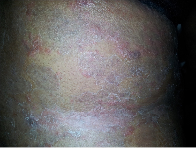
PLATE 4: Tinea pedis in an
HIV seropositive patient

PLATE 5: Tinea corporis in a
young HIV seropositive male
MYCOLOGY:
(1) Potassium Hydroxide Wet Mount:
Eleven
of the specimens from the HIV sero-positive cases (91.6%) were positive for
fungal hyphae while 4 of the control group(80%) were
positive for fungal hyphae.
(2) Culture:
Five
of the specimens from the HIV sero-positive cases (41.6%) grew
dermatophytes.Out of this number, 2 (16.6%) were Trichophyton mentagrophyte; 2(16.6%) were Trichophyton
soudanenses and 1(8.3%) was Microsporum auodunii. Two of the
specimens (16.6%) showed no growth, while 5(41.6%) specimens grew
non-dermatophytic fungi such as Aspergillus
fumigatus, Aspergillus niger, and Penicillium chrysogenum.
For the control, 2 (40%) of the specimen
showed no significant growth, while 3 of the specimens grew non-dermatophytic
fungi (Aspergillus niger,
Aspergillus fumigatus and Aspergillus flavus). Other details are in the
tables below.
TABLE 12: MYCOLOGY
RESULTS OF HIV POSITIVE CASES WITH CLINICALLY DIAGNOSED DERMATOPHYTOSIS
|
SPECIES
|
FREQUENCY
|
PERCENTAGE
|
|
Microsporum auodonii
|
1
|
8.3%
|
|
Trichophyton
soudanenses
|
2
|
16.6%
|
|
Trichophyton
mentagrophyte
|
2
|
16.6%
|
|
Other
species (Aspergillus,penicillium)
|
5
|
41.6%
|
|
No
significant growth
|
2
|
16.6%
|
|
Total
|
12
|
100%
|
TABLE 13: MYCOLOGY
RESULTS OF THE CONTROLS WITH CLINICALLY DIAGNOSED DERMATOPHYTOSIS
|
SPECIES
|
FREQUENCY
|
PERCENTAGE
|
|
Non-
dermatophytic fungi
|
3
|
60%
|
|
No
significant growth
|
2
|
40%
|
|
TOTAL
|
5
|
100%
|
PLATES 7-9: Dermatophytes cultured from the specimens collected from the subjects

PLATE
7:
Culture of Microsporum audounii in
Saboraoud's dextrose agar (SDA).

PLATE 8: Culture of Trichophyton mentagrophyte in SDA.

PLATE
9: Culture of Trichophyton soudanense in SDA.
DISCUSSION
This study was
carried out to assess the prevalence and pattern of the various types of
dermatophytosis among HIV infected patients as compared to apparently healthy
seronegative controls.
Various skin conditions are associated with HIV infection. Epidemiologic
studies have shown that almost all persons with HIV infection will have skin
disorders at some point during their disease.96 Skin disorders commonly encountered in HIV-infected
patients may be the first manifestation of HIV disease. Up to 90% of
HIV-infected persons suffer from skin diseases during the course their of illness97. In a recent cross-sectional
study of 186 HIV positive patients, 175 (94%) suffered from one or more
cutaneous disorders98. The most common skin disorder
identified was fungal infection, followed by eczema and seborrhoeic dermatitis.
The spectrum of skin disorders depends on: (a) immunologic stage, as reflected
by CD4 count
(b) concurrent use of Highly Active Anti-Retroviral Therapy (HAART) (c) pattern
of endemic infections.
In general,
declining immunity is associated with increased number and severity of skin
disorders99. Skin
lesions are more likely to have unusual appearances in advanced HIV infection.
In this study the prevalence of several skin diseases was found to be
significantly higher in the HIV seropositive group compared to the seronegative
controls (34.6% vs 6.9%).The mean duration of the lesions was also longer for
the cases compared to the control group (71 weeks vs 8weeks). In addition, the
mean CD4 cell count of the HIV seropositive cases with skin disease was
significantly lower than those without any skin disease thus
indicating the importance of immunosuppression in the development of skin
disease in such patients.
Dermatophytosis
is the second commonest skin lesion in the HIV seropositive group (next to
pruritic papular eruption of HIV). It has a significantly higher prevalence
among the cases when compared to the control group -6.9% vs2.8%, (p<0.05). The prevalence of dermatophytosis in the HIV
positive cases used in this study is 6.9%. Previous studies done in the past
revealed prevalence rates between 6.06% and 30%50-52,70,100-102.
The relatively low prevalence observed in this study may be
attributable to the relatively fewer number of subjects studied and to the low
frequency of contact with infected persons as observed in this study (only 2.8%
of cases admitted to having any history of contact with other persons with skin
lesions).
Cases with
dermatophytosis have a mean CD4 cell count of
226.2cells/µl, this figure can be said to be much lower when compared to
other studies where mean CD4 cell counts
in HIV positive patients with dermatophytosis ranged from 267-450 cells/µl51,103,104. Of the cases with clinical dermatophytosis,
41.6% % have CD4 cell count between 0 and 200 cells/µl.
From these findings it can be concluded that dermatophytosis is directly
related to the degree of immunosuppression in HIV sero-positive patients.
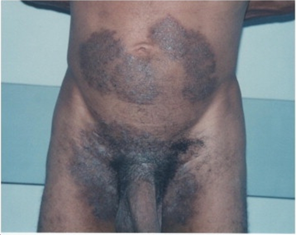
Tinea corporis
is the commonest dermatophytic lesion seen among the cases. In previous studies sited earlier
it was also found to be the commonest in the setting of HIV infection. This is
in keeping with other studies where tinea corporis or capitis where found to be
the commonest dermatophytosis affecting HIV patients.70,97,102,105 All of the cases seen in this study had a CD4
cell count below 400cells/µl (two thirds
below 200 cells/µl). Other studies done in the past revealed a similar high prevalence in
patients with low CD4 cell count. Most of the lesions seen were of the classical annular types with
active edges and healing centers or tinea incognito. A female among the control
with tinea incognito admitted to chronic application of bleaching creams (see
plate 4). Two of
the cases with tinea corporis had extensive involvement of the trunk, limbs and
flexures, with hyperpigmented, thick scaly plaques (see plate 6). This is in
keeping with findings
from previous studies that have demonstrated atypical presentations of tinea
corporis in immunosuppressed persons with HIV infection. The paucity of such
atypical lesions (in this study) described in earlier literature72-74, may be attributed
to the fact that a significant number of the patients(16.1% of the cases) have
utilized one form of antifungal treatment or another prior to presentation.
Another common
type of dermatophytosis seen among the cases was tinea unguium, the patients with these lesions had a CD4
count of 400
cells/µl and below. Moreover, all of the cases with onychomycosis fall
within the 21-40 year age group who are more prone to trauma compared to other
age groups (prior trauma is a risk factor for onychomycosis106. It
is rare in HIV positive children. In a recent study on prevalence of
dermatophytosis in HIV positive children in Nigeria, Tinea unguium constituted just 5% of the total
no of dermatophytosis seen in the entire population.105
The typical lesions
seen in HIV- proximal
white subungual onychomycosis and superficial white onychomycosis
were not seen in this study. However, the patients seen had nail dystrophy,
discolouration, onycholysis and nail destruction (see
plate 2). Onychomycosis in the setting of HIV infection usually involves the
toe nails, however in this study, the cases seen were in the finger
nails. This may be attributed to the fact that most of the cases seen were
women (61.2 %) who
are more exposed to moisture in the course of their household chores than their
male counter parts.
Tinea capitis, a
common dermatophyte infection in children was not seen in any HIV positive
child. This may be attributed to the fact that only a small proportion of
children were used in this study. A 13 year old school boy in the control group
had it. He presented with patchy alopecia.
Tinea manuum and tinea pedis were encountered less
frequently in this study(16.6% each) and the
morphology of the lesions seen were not significantly different from those seen
in immunocompetent HIV negative persons. No case of tinea cruris was seen in
this study. This may be attributed to the fact that a relatively fewer number
of males were used in this study since it occurs more frequently in the male
sex.107
There is a low mycology yield of specimens cultured in
this study. This may be attributed to adulteration of the culture media or
prior treatment of the skin lesions by the subjects.
The commonest
dermatophyte isolated in this study was trichophyton species (33.2% of
isolates) This is in keeping with findings in several other studies where it
has been found to be the commonest aetiologic agent of dermatophytosis in HIV
infected patients. 17,102,108,109
Microsporum species was another common dermatophyte isolated
(8.3%). This is also a common dermatophyte seen in HIV patients from previous
studies97,110. It occurs in severe
immunosuppression and can be invasive.
Aspergillus species was isolated in some (41.6%) of
the HIV infected patients with dermatophytosis. However, this organism is not
commonly associated with dermatomycosis in HIV. It may be an incidental finding
or a contaminant.111
Penicillium
species were also isolated in some(41.6%) of the
patients. These may either be due to the contamination,
increased susceptibility to non-dermatophyte infections in HIV infected persons
due to immunosuppression or environmental factors that favour the growth of
non-dermatophytic fungi.
CONCLUSION
This study was
carried out to determine the prevalence and clinical variations of the various
types of dermatophytosis among HIV seropositive patients seen in Portharcourt,
Southern Nigeria.
A significantly
higher prevalence of dermatophytosis as well as other skin lesions was observed
in the HIV positive cases when compared to the control and most of the patients
had advanced HIV infection as evidenced by a low CD4 cell count (below
200cells/µl). Thus it can be concluded that the occurrence of dermatophytosis
in HIV infected persons is positively associated with the degree of
immunosuppression.
The age group with the highest prevalence of
dermatophytosis is the 21-40 and 41-60 year age group. Males have a higher prevalence than females.
The commonest lesion
found in this study was tinea corporis. Other dermatophyte lesions seen were
tinea capitis, tinea manuum , pedis and
onychomycosis..
The various atypical dermatophytic lesions
found in HIV infected persons as reported in other studies were not commonly
seen. Most of the skin lesions seen in this study were not much different from
the classical lesions seen in HIV seronegative persons with the exception of 2
cases of disseminated and atypical presentation of tinea corporis. Proximal
white subungual onychomycosis, said to be pathognomonic of HIV/AIDS was not seen
in any patient enrolled for this study.
The commonest
aetiologic agents for dermatophytosis in the group of HIV infected patients
studied are trichophyton species and microsporum species which are known to be
common aetiologic agents of dermatophytosis in HIV infected patients from
previous studies. Other fungi that were commonly isolated include aspergillus
species, and penicillium species but these are of doubtful significance.
Acknowledgement
Dr Eli Sukarime
Executive Director Mother, Baby and
Adolescent Care Global Foundation ( Previously Mother
and Baby Care Global Foundation (April 2021)
REFERENCES
(1.) Christine MP.
Geographical distribution of the dermatophytes: A review. J. Hyg. Camb. 1978. 80: 301-310.
(2.) Nweze EI, Okafor
JI. Increasing cases of tinea capitis dermatophytosis in children of school
age in Nigeria. (2007). Second Pan African medical mycology
society conference, Cape Town. South Africa.
(3.) Odunowo DB, Pattern of Dermatophytosis among
out-patients seen in Lagos university teaching hospital skin clinic. (1988) pg92.
(4.)
Jacobs PH. Dermatophytes that infect animals and humans. Cutis, 1988;
42:130-131.
(5.) Olumide YM. A pictorial self-instructional
manual on common skin diseases (where there is no dermatologist). College of Medicine of the University of Lagos Press 1990.15-23.
(6.)Mackenzie DWR. "Hairbrush diagnosis" in detection
and eradication of non-fluorescent scalp ringworm. British
medical Journal 1963; 2: 363-365.
(7.) Rothman S, Smiljanic L, Shapiro L, Weitkamp AW. The spontaneous cure of tinea capitis in puberty. J. Invest.
Dermatol. 1947. 8: 81-98.
(8.) Calderon RA,
Hay RJ. Fungicidal activity of human neutrophils and
monocytes on dermatophyte fungi, Trichophyton
quinckeanum and Trichophyton rubrum.
Immunology 1986; 61: 289–95.)
(9.) Hay RJ, Reid
S, Talwat E, Macnamara K. Immune responses of patients with tinea imbricata. Br J Dermatol 1983; 108: 581–9.
(10.)
Calderon RA, Hay RJ. Cell-mediated
immunity in experimental murine dermatophytosis.II. Adoptive
transfer of immunity to dermatophyte infection by lymphoid cells from donors
with acute or chronic infections. Immunology 1984;
53: 405–10.
(11.) Jones HE,
Reinhardt JH, Rinaldi MG. Acquired immunity to dermatophytosis. Arch Dermatol 1974; 109: 840–8.
(12.) Hay RJ, Shennan G. Chronic dermatophyte
infections. II. Antibody and cell mediated immune responses. Br
J Dermatol. 1982; 106: 191–5.
(13.) Serjeantson S, Lawrence G. Autosomal recessive inheritance of
susceptibility to tinea imbricata. Lancet 1977; 1: 13–15.
(14.) Gilchrest BA. Some
gerontologic considerations in the practice of dermatology. Arch.
Dermatol.1979;155:1343-1346.
(15.) Hay RJ. Chronic dermatophyte
infections. I. Clinical and mycological features. Br J
Dermatol 1982; 106: 1–6.
(16.) Canizares O, Shatin H, Kellert AJ. Cushing,s syndrome and dermatomycosis. Arch.
Dermatol.1959.80:705-715.
(17.) Torssander
J, Karlsson A, Morfeldt-Månson L, Putkonen PO, Wasserman J. Dermatophytosis
in HIV infection: a study in homosexual men. Acta Derm.
Venereal (Stockti) 1998; 68: 53 – 56.
(18.) Allen AM, King RD. Occlusion, carbon dioxide and fungal skin
infections. Lancet1978; 1: 360–2.
(19.) Allan Ll, Sung
Huang S. Dermatophyte Viability at Modestly Raised Temperatures Arch Dermatol.
1963; 88(4):393-402.
(20.) CDC (Centre for Disease Control) (1981c)
follow up on Kaposi Sarcoma and pneumocystic pneumonia. MMWR
(Morbidity and Mortality weekly Report) 1981; 30; 409 – 410.
(21.)
Centre For Disease Control (1981) Kaposi Sarcoma and
pneumocystic pneumonia among homosexual men – New York City and California
MMWR. 1981; 30: 305-308
(22.)
Centre For Disease Control (1981a). Pneumocystic
pneumonia – Los Angeles MMWR 1981; 30: 250 – 252.
(23.) Barré-Sinoussi F, Chermann JC, Rey F, Nugeyre MT, Chamaret S, Gruest J, et al .
Isolation of a T-Lymphotrophic retrovirus from a patient at
risk for AIDS. Science 1983, 220: 868 – 871.
(24.)
Broder S, Gallo RC, A pathogenic retrovirus (HTLV III). Linked
to AIDS. New England Journal of Medicine 1984, 311: 1292 – 1297.
(25.)
Gallo RC, Salahuddin SZ, Popovic M, Shearer GM, Kaplan M, Haynes BF, et al.
Frequent detection and isolation of cytopathic retroviruses (HTLV –
III) from patients with AIDS and at risk for AIDS. Science 1984, 224:
500 – 503.
(26.) UNAIDS Report on the global AIDS
epidemic | 2020.
(27.)National Agency for the Control of AIDS (NACA). Federal Republic of Nigeria. Global AIDS
response. Country Progress Report. Nigeria GARPR. 2012. P 16-17.
(28.)
Braathen LR, Kaanan T. Human epidermal langerhan’s cells induce cellular immune
responses to trichopyton in dermatophytosis. Br. J. Dermatol. 1983; 109; 295 – 9.
(29.)
Groh V, Mann DL. Langerhan’s cells in HIV-I infection.
J. Am. Acad. Dermatol., June 1996; 134(6) 1122 – 1124.
(30.)
Stingl G, Rappersberger
K, Tschachler E, Gartner S, Groh V, Mann DL, et
al. Langerhan cell in HIV I infection. J. Am. Acad. Dermatol. 1990; 22: 1210 – 1217.
(31.) Reeves, J. D. Doms, R. W. "Human Immunodeficiency
Virus Type 2". J. Gen. Virol.
2002. 83 (Pt 6): 1253–65.
(32.)Thomson MM, Perez-Alvarez L,
Najera R (2002). "Molecular epidemiology of HIV-1
genetic forms and its significance for vaccine development and therapy".
Lancet Infectious
Diseases 2002. (8): 461–471.
(33.) Carr
JK, Foley BT, Leitner T, Salminen M, Korber B, McCutchan F. "Reference sequences
representing the principal genetic diversity of HIV-1 in the pandemic" (PDF). In Los Alamos
National Laboratory (ed.). HIV sequence compendium. Los Alamos, New
Mexico:
Los Alamos National Laboratory. 1998. pp. 10–19.
(34.) Markowitz, edited by William N. Rom ; associate editor, Steven B. Environmental
and occupational medicine (4th ed.). Philadelphia: Wolters Kluwer/Lippincott
Williams & Wilkins. . 2007. p. 745.
(35.)
Boily MC, Baggaley RF, Wang L, Masse B, White RG, Hayes RJ, Alary M . "Heterosexual risk of HIV-1 infection
per sexual act: systematic review and meta-analysis of observational
studies". The
Lancet Infectious Diseases 2009.
(2): 118–129.)
(36.) HIV in the United States: An
Overview".
Center for Disease
Control and Prevention. March 2012.
(37.) Fiona Samuels and Sanju Wagle 2011. Population mobility and HIV and AIDS:
review of laws, policies and treaties between Bangladesh, Nepal and India. London: Overseas Development Institute).
(38.) Coutsoudis A, Kwaan L,
Thomson M."Prevention of vertical transmission of HIV-1 in
resource-limited settings". Expert
review of anti-infective therapy 8
.2010 Oct (10): 1163–75.
(39.) Baggaley, RF; Boily,
MC; White, RG; Alary, M. "Risk of HIV-1 transmission for parenteral
exposure and blood transfusion: a systematic review and meta-analysis". AIDS (London, England) 2006: 20 (6): 805–12).
(40.)
Kripke, C . "Antiretroviral
prophylaxis for occupational exposure to HIV.". American family physician 2007.76 (3): 375–6.
(41.) Parren PW, Moore J, Burton D, Sattentau Q. The neutralising
antibody response to HIV-1: viral evasion and escape from humoral immunity. AIDS
1999; 13:
S137–S162.
(42.) Wilkinson J, Gotch FM. Immune interventions. Br Med Bull 2001; 58:
187–203.
(43.) Meinl E, Fickenscher H, Thome M, Tschopp J, Fleckenstein B. Anti-apoptotic
strategies of lymphotropic viruses. Immunol. Today 1998; 19: 474–9.
(44.) Veazey R, Lackner A. The mucosal immune system
and HIV.1 infection. AIDS Rev.
2003; 5: 245–52.
(45.) World Health Organization. WHO Case Definitions of HIV for Surveillance and
Revised Clinical Staging and Immunological Classification of HIV-Related
Disease in Adults and Children; 2007.
(46.)
Fauci AS, Clifford Lane H. HIV disease, AIDS and related disorders. In
Braunwald E, Hanser SL, Fauci AS, Long DL, Kasper DL, Jameson JL, Editors,
Harrison’s Principle of Internal Medicine 15th Edition. New York
Mc-Graw Hill Vol. 2 P. 1852 – 1912.
(47.)
Olumide YM, Dada AJ, Sogbanmu TB, Aruna GB. Seroprevalence study of HIV I, HIV
II & HTLV I among patients at the Dermatovenerology Clinic of the Lagos
University Teaching Hospital. Int. J. Dermatol. 1999 Oct; 36 (10): 741 – 4.
(48.)
Yahaya H. Change in pattern of skin disease in Kaduna, North Central Nigeria.
Int. J. Dermatol. 2007 Sept; 46(9): 936 – 43.
(49.)
Ogunbiyi AO, Daramola OO, Alese OO. Prevalence of skin diseases
in Ibadan Nigeria. Int. J. Dermatol. 2004 Jan. 43(1): 31 – 6.
(50.) Goodman DS, Teplitz ED, Wishner A, Klein RS, Burk PG, Hershenbaum E. Prevalence of
cutaneous disease in patients with AIDS or AIDS Related Complex. J. Am. Acad.
Dermatology. 1987; 17:210 – 220.
(51.)
Nnoruka EN, Chukwuka JC, Anisiuba B. Correlation of mucocutaneous manifestation
of HIV/AIDS infection with CD4 Counts and disease progression. Int.J. Dermatol.
2007 Oct. 46 Suppl. 2, 14–8.
(52.)
Shobhana A, Guha SK, Neogi, DK. Mucocutaneous manifestations of HIV infection. Indian Journal of Dermatology Venerology and Leprology. Vol. 70; No 2. March – April 2004. Pp 82-86
(53.) Aly R, Berger T. Common Superficial fungal
infections in patients with AIDS. Clinical Infectious
Disease. 1996; 22: 5128 – 5132.
(54.)
Ajao AO, Akintunde C. Studies on the prevalence of tinea capitis infection in
Ile-Ife, Nigeria. Mycopathologica 1985, Jan;89(1):
43-8.
(55.)
Ameh IG, Okolo RU. Dermatophytosis among school children in Nigeria: Domestic
animals as predisposing factors in Sokoto, Nigeria. Pakistan Journal of
biological sciences 2004 (7); 1109-1112..
(56.)
Sanuth HA, Efuntoye M.O. Distribution and microbiological characteristics of
dermatophyte infection among primary school children in Ago-Iwoye, Oguun state,
Nigeria. Researcher. 2010: 2(12): 80-85.
(57.)
Ndako JA, Osemwegie OO, Spencer TH, Olopade BK, Yunusa GA, Banda J. Prevalence
of dermatophytosis and other associated fungi among school children. Global
advanced research journal of medicine and medical sciences. Vol.
1(3) pp.049-056, April 2012.
(58.)
Akinboro AO, Olasode AO, Onayemi O. The pattern, risk factors
and clinico-aetiological correlates of tinea capitis among the children in a
tropical community setting of Osogbo, Southwestern Nigeria. Afro-Egypt
J. Infect. Endem. Dis. 2011, 1(2): 53-64.
(59.)
David DL, Edward A, Zaruwa MZ, Addas PA. Barbing salon associated fungal
disease infection in Mubi, Adamawa state, Nigeria. World journal of medical
sciences, 5(1): 17-21, 2010.
(60.)
Soyinka F. Epidemiological study of dermatophyte infections in Nigeria
(clinical survey and laboratory investigations). Mycopathologia
1978. July 28; 63 (2) 99-103. (61.) Zurita J, Hay RJ. The
adherence of dermatophyte microconidia and arthroconidia to human keratinocytes
in vitro. J. Invest, Dermatol. (1987); 89: 529-534.
(62.)
Al Jabre SH, Richardson MD, Scott EM, Rashid A, Shankland GS. Adherence of arthroconidia and
germlings of anthropophilic and zoophilic varieties of Trichophyton mentagrophytes to human corneocytes as an early event
in the pathogenesis of dermatophytosis. Clin Exp
Dermatol 1993; 18: 231–5.
(63.) Blank H, Sagami S, Boyd C, Roth FJ. The pathogenesis of
superficial fungal infections in cultured human skin. Archives of Dermatology, 1959;
72: 524-535.
(64.) Lorincz AL, Priestly JO, Jacobs PH. Evidence for a humoral
mechanism which prevents growth of dermatophytes. Journal of Investigative
Dermatology, 1958; 31:15-17.
(65.) Raubitschek F, Maoz
R. Invasion of nails in vitro by certain dermatophytes. J. Invest Dermatol
1957; 28: 261–8.
(66.) Lambkin E, Hamilton A, Hay RJ. Partial purification and
characterization of a 235 000 Mr extracellular proteinase from Trichophyton rubrum. Mycoses 1994;
37: 85–92.
(67.) Yu RJ, Harmon SR, Grappel SF. Two cell
bound keratinases of Trichophyton
mentagrophytes. J Invest Dermatol 1971; 56: 27–32.
(68.) Siesenop U, Bohm KH. Comparative studies
on keratinase production of Trichophyton
mentagrophytes of animal origin. Mycoses 1995; 38:
205–9.
(69.) English MP. Nails and fungi. Br
J Dermatol 1976; 74:
697–701
(70.) Kaviarasan PK, Jaisankar TJ, Thappa DM, Sujatha S. Clinical
Variations in Dermatophytosis in HIV infected patients. Indian Journal of
Dermatology, Venerology and Leprology 2002, 68: 213 – 216.
(71.) Ekong
, A. Uwa, and I. Grant – Prevalence and types of opportunistic
infections in HIV/AIDS patients in Nigeria. 5th International
Congress on Drug Therapy in HIV infection. 22 – 26 October
2000 Glasgow UK.
(72.)
Smith KJ, Skelton HG, Yeager J, Ledsky R, McCarthy W, Baxter D, et al, Cutaneous findings in HIV I positive
patients a 42 month prospective study. Military Medical Consortium for the
advancement of retroviral research (MMCARR) J. Am. Acad. Dermatol. 1994; 31: 746 – 754.
(73.) Berger TG, Greene I. Bacterial, Viral, Fungal
and Parasitic infections in HIV disease and AIDS. Dermatol.
Clin. 1991; 9: 465 – 492.
(74.) Muñoz-Pèrez MA, Rodriguez-Pichardo A, Camacho F, Rios JJ. Extensive and deep
dermatophytosis caused by Trichophyton
Mentagrophytes var interdigitale in an HIV – I positive patient. Journal of
the European Academy of Dermatology and Venerology .
2000.14 (1): 61 – 63.
(75.)
Kwon KS, Jang HS, Son HS, Oh CK, Kwon YW, Kim KH, et al.
Widespread and invasive Trichophyton
Rubrum infection mimicking Kaposi
Sarcoma in a patient with AIDS. J. Dermatol. 2004. October; 31 (10): 839 – 843.
(76.)
Vilela R, Oliveira TF, Vilela L,
Mendoza L. Exacerbated
inflammatory reaction to Trichophyton rubrum infection on an
HIV-positive patient successfully treated with fluconazole Mycopathologica Vol.
163. No 4, April 2007 page 208 – 205.
(77.) Porro AM, Yoshioka MC, Kaminski SK, Palmeira Mdo C, Fischman O, Alchorne MM. Disseminated
dermatophytosis caused by Microsporum
gypseum in 2 patients with AIDS. Mycopathologica 1997; 137 (1) 9 – 12.
(78.)
Nenoff P, Graser Y, Kibuka – Serunkuma
L, Muylowa, GK. Tinea circinata manus due to microsporum gypseum in an HIV positive
boy in Uganda East Africa. Mycosis. Vol. 50; No
2 March 2007, Pp 1 53 – 155.
(79.)
Nuniman C, Fernandez, Fabriciolamy, Tiyomi Akiti – Microsporum gypseum infection in AIDS patient. A
case report. Anais brasileires de dermatologica. Vol.
73 No 1, (Jan/Feb 1998).
(80.) Menon T, Ranganathan S. Extensive T. Corporis caused by Trichophyton verrucosum in a patient
with HIV infection. Indian Journal of Dermatol. Venerol and Leprol. 1996; 62: 126 – 127.
(81.) Lowinger Seoane M, Torres-Rodriguez JM,
Madrenys-Brune N, Aregall-Fuste S, Saballs P.
Extensive dermatophytosis caused by Trichophyton
Mentagrophytes and Microsporum Canis in
a patient with AIDS. Mycopathologica, 1992 Dec. 120 (3): 143 – 146.
(82.) Rothman S, Smiljame A, Shapiro AL, Weltkamp
AW, The spontaneous cure of Tinea capitis at puberty. J. Invest. Dermatol. 1947; 8: 81 – 98.
(83.)
Tinea Capitis in adults and adolescents. Arch. Dermatol. Syphilol.
1952; 65: 9 – 40.
(84.) Gremer G, Bournerias I, Vandeleme-Ubroucke E,
Hourn R, Revur J, Tinea Capitis in adults – misdiagnosis or reappearance. Dermatology 1997; 194: 8 – 11.
(85.) Lateur N, Andre J, De Maubeuge J, Poncin M,
Song M, Tinea Capitis in 2 black African adults with HIV infection. Br. J.
Dermatol. 1999 April; 140(4): 722 – 724.
(86.)
Bournias I, Dechauwin MF, Datry A, Chambrette I, Camiere J, Deridas A, et al.
Unusual microsporum caris infections in adult HIV infected patients. J. Am.
Acad. Dermatol. 1996 Nov.; 35 (5p+2) 808 – 810.
(87.)
Gregory N. Special patient populations: onychomycosis in the HIV positive
patient. J. Am. Acad. Dermatol. 1996; 35: 813 – 816.
(88.)
Levy LA, Epidemiology of onychomycosis in special risk populations. J. Am. Paediatric Medical Association. 1997;
87: 546 – 550.
(89.)
Dompmartin D, Dompmartin
A, Deluol AM, Grosshans E, Coulaud JP. Onychomycosis in AIDS - Clinical and laboratory
findings in 62 patients. Int. J. of Dermatol. 1990
June, 29 (5) 337 – 339.
(90.)
Odom RB. Common
superficial fungal infections in immunosuppressed patients. J. Am. Acad. Dermatology 1994; 31: 856 – 859.
(91.) Daniel CR, Norton LA, Scher RK. The Spectrum of nail disease in patients with HIV. J. Am. Acad. Dermatol 1992; 27: 93 – 97.
(92.)
Raninborg L,
Baastrup N, Svejguard E. Onychomycosis in HIV infected patients. Acta Dermatol. Venerol. 1998; 78: 151 – 152.
(93.)
Svejgaard E. Recent trends in the immunology of dermatophytes. Microbial. Sci. 1986; 3: 154 – 159.
(94.)
Popoola DA, Ojo DA,
Alabi RO, Prevalence of dermatophytosis in junior secondary
school children in Ogun state Nigeria. Mycoses. 2006.
Vol.49 issue 6.:499-502.
(95.) Ozumba UC, Nlemadim R. Prevalence of
dermatophytosis in University of Nigeria Teaching Hospital, Enugu, Nigeria. Any
change in pattern. Niger. J. Clin.Pract. 2005.
Dec. 8(2):83-88.
(96.)
Coldrion BM,
Bergshesser PR. Prevalence and clinical
spectrum of skin disease in patients infected with human immunodeficiency
virus. Arch. Dermatol. 1989. 125:357-61.
(97.)
Pennys NS. Skin manifestation of AIDS. London Martin. Dumitz 1995.
(98)
Ho KM, Wong KH.
Dermatological Manifestations in HIV disease. In Chan
KLW, Wong KH, Lee SS, eds. HIV manual 2001.pp 231-245.
(99.) Raju PV, Rao GR, Ramari TV, Vandana S.
Skin disease: Clinical indicator of immunostatus in human immunodeficiency
virus (HIV) infection. Int J. Dermatol.2005; 44:646-9.
(100.) Kumarasamy N, Solomon S, Madhivanan P, Ravikumar B, Thyagarajan
SP, Yesudian P. Dermatologic manifestations among human
immunodeficiency virus patients in South India. Int J. Dermatol.2000:
39:192-195.
(101.) Singh A, Thappa DM, Hamide A. The
spectrum of mucocutaneous manifestations during the evolutionary phases of HIV
disease. An emerging Indian scenario. J.
Dermatol. (Tokyo)1999;26: 294-304.
(102.)
Rajesh R, Subramanian K, Padmavasthy BK, Vasanthi S. Prevalence and specie
profile of dermatophytosis among HIV positive patients in a rural referral
centre. Indian J. Sex. Transm.
Dis. 2006; vol. 27 No 2p70.
(103.)
Satiya VSA, Singh VP, Shyam S, Gulati AK, Varma DV, Rai M. Relationship between
skin diseases and cd4 cell count in a hospital based cohort of HIV infected adults in North India.Journal
,Indian Academy of clinical medicine. March 2008. Vol 9, No 1Jan- p 20-25.
(104) Conant MA. The AIDS Epidemic.
J
Am Acad Dermatol 1994 Sep;31(3pt2):47-50.
(105.) Umoru DD,
Esene H. Clinical characteristics of
dermatophytosis among children in a Nigeria population; the role of HIV/AIDS
Benin journal of postgraduate medicine. vol 12 no 1 Dec.
2010. p 32-36.
(106.) Daniel CR III, Gupta AK, Daniel MP, Daniel CM. Two feet-one hand
syndrome: a retrospective multicenter survey. Int J Dermatol. 1997;36:658-660
(107.)Silva-Tavares H, Alchorne MM, Fischman O. Tinea cruris
epidemiology (São Paulo, Brazil). Mycopathologia. 2001;149:147-149.)
(108.) Adriano S, Valeri B, Giuliano B, Gingio B,
Anna M, Severino S. Prevalence of dermatophytosis and yeasts in HIV patients .
Mycopathologia
vol. 14, No 2, 103-107,d01.1007/BF0043629.
(109.)
Amar S, Ratnakar K ,Chetan O, Dattatray S, Minal T, Sujata T et al. A clinical and mycological study of onychomycosis in HIV infection
Indian J. Dermatol.
Venereal. Leprol. 2007 397-401.
(110) Bakos L, Bonamigo RR. Pisani AC.
Mariante JC, Mallman R. Scutular favus-like tinea cruris and pedis in a patient
with AIDS. J Am Acad Dermatol 1996;34:1086-7.
(111.) Primary cutaneous
aspergillosis in Human Immunodeficiency virus -infected patients. Clinical
infectious Diseases. Sep 1998. Vol. 27. No
3:p641-643.
|
Cite this
Article: Amaewhule MN (2021). Prevalence and Pattern of Dermatophytosis
in Patients with Human Immunodeficiency Virus Infection Seen in The
University of Port Harcourt Teaching Hospital, (UPTH) Port- Harcourt. Greener
Journal of Medical Sciences, 11(1): 46-72.
|