Identification
of fungal pathogens associated with mango disease in UsmanuDanfodiyo University, Sokoto
was carried out in selected Areas of the University, with the aim to Isolate and identify the fungi associated with fruits, leaves and
stem, and genotypically characterize the most
significantly occurred fungal isolates by PCR assays. Mango
trees were selected from four different locations; there are about eight
different symptoms associated with the diseases of mango trees in the study
site. These include; gum exudation, bark splitting, discoloration and
darkening of the bark, wilting of leaves, root decay, whitish substance on
and around the root surfaces and wilting of branches. Based on morphological identification, 448 fungal isolates were
tentatively identified into 7 genera and 11 species, namely Alternariaalternata,
Aspergillusflavus,
A. fumigatus, A. niger,
Colletotrichumgloeosporioides,
Fusariumsemitectum, Papulasporasp., Penicilliumsp., Pestalotiopsisguepiniiand Rhizopusstoloniferamong others. The
extracted DNA from the most frequent selected isolates of fungal species was
successfully amplified with universal primer ITS1 and ITS4 primer,
sequencing confirmed the identification of 7 species of fungi from gel amplicons with 99-100% similarities index. The present
study showed that diverse groups of fungal genera were associated with leaf
spot of mango. The F. verticillioidesand L. theobromaewere found to
be associated with anthracnose of mango, implying that other fungal species
may act in synergy with C. gloeosporioidesto
cause fungal disease of mango in the study areas. The
study recommends awareness creation on good orchard management practices,
regular monitoring of the orchards by the management and establishing a link
with an expert for advice.
Accepted:29/08/2023
Published: 09/09/2023
*Corresponding
Author
Ibrahim Alhaji SALAU
E-mail: ibrahimasalau@yahoo.com
Phone: +234(0) 8053569551
Keywords: Fungi,
disease, Mango, Molecular, UDUS.
INTRODUCTION
Mango (MangiferaindicaL.) is the most popular and commonly eaten fruit
among millions of people in tropical areas; it is a family member of the Anacardiaceae, one of the crops grown abundantly
throughout the Nigerian tropics. The cultivars of mango commonly found in
Nigeria are Edward, Early Gold, Local Alphonso, July
Haden and Ogbomosho (Okibo 2001). The crop is a host
to a large number of pathogens such as bacteria, fungi and viruses (Kumar et
al., 2007). Fungal pathogens are frequently encountered on leafs, stem and
rotting mango fruits, and are the major agents of rot after harvest (Diedhiouet al., 2007). Mango tree vegetation in UsmanuDanfodiyo University, Sokoto is under threat by identified and unidentified
pathogens. Diseases has rendered its production non attractive to both farmers
and gardeners in the university. So far many works were made by researchers on
mango fruits collected from different parts of the state, but little or no
attention is paid to foliar and stems diseases of the tree.
Studies from several parts of the world where mango is
grown have shown that fungi are the most devastating disease which does not
only reduce mango fruit yield but also render marketable fruits worthless. In
Nigeria, mango production and exportation is greatly limited due to post
harvest rot of fruits associated with fungi and over 30% of harvestable fruits
are lost annually because of fruit abortions and abscission caused by fungi (Onyeaniet al. 2012).Like any other crops, mango is
also susceptible to diseases caused mainly by Ascomycetes
and mitosporic fungi. One of the major diseases of
mango is leaf spot, caused by the fungi from the genera Colletotrichum,
Alternaria, Cercospora,
Curvularia, Cladosporium,
Ascochyta and Botryodiplodia(Agrios, 2005). Symptoms of leaf spot can vary
depending on the fungal pathogen. The spot vary in size and shape but commonly
begins with pinhead point’s lesion and spread forming circular or irregular
lesion with dry, brown or black raised centre. The
infection of pathogen will cause chlorosis and
necrosis on the leaf surface and thus reduce photosynthetic areas, which
affects carbohydrate production as well as nutrient transportation to plant
organs (Agrios, 2005). Consequently, the infection
will reduce plant growth and fruit yield.
As many fungal genera can cause leaf spot disease,
identification of the causal pathogen is important to initiate preventive or
curative measures. For that reason, accurate identification of fungal pathogens
is necessary to determine appropriate disease control measures as well as to
improve disease management. The most prevalent technique used to identify plant
pathogens is by observing morphological characters. Morphological
characteristic commonly used for identification of fungi, include macroscopic
and microscopic characteristics such as colony colour
and texture, pigmentation, growth diameter, the shape of conidia, arrangement
of spore or conidia, conidiophore, presence of resistant structure such as chlamydospore and presence of fruiting bodies such as pycnidia and acervuli (Watanabe,
2002; Barnett and Hunter, 2006).
In Sokoto state, fungal species
associated with mango leaf is not well-documented and its Pathogenicity has not
been reported. Therefore, identification and characterization of fungi causing
fruits, leaf and stem disease of mango based on morphological and molecular
approaches are important in order to protect the plant from further damage as
the yield can be affected.
In the last few decades, molecular approaches employing
the Polymerase Chain Reaction (PCR) has been found to provide sensitive means
of characterizing and classifying plant pathogenic fungal species (Etebu and Osborn, 2012). In this study, molecular approach
was adopted in a view to using region of DNA sequence best suitable for
accurate identification of the fungal pathogens associated with mango diseases.
MATERIALS
AND METHODS
Study Area
The research was carried out at UsmanuDanfodiyo University Sokoto
permanent site in Wamako Local Government Area of Sokoto State which is at the extreme of north western block
of Nigeria between latitude 11.60N-13.90N and longitude
3.70E-6.90E (Bello, 2002). The area falls under Sudan
savanna zone characterized by two distinct seasons (wet and dry) of varying
duration and intensity (Anon, 2014). The rainy season is often 3-4 month
(usually from May to September) with the highest rainfall in August, the mean
annual rainfall 500-750mm with, relatively high temperature though it varies
with season (Anon, 2014).
Sampling Technique
Purposive sampling was employed to
sample out the diseased trees in the study area as the whole area was divided
into four (4) locations; each location was further sub-divided into four (4) blocks to serve as a replicate. The locations were:
Location 1: This extends from Kofar Mata through Female hostel, behind Convocation Arena,
and the Male hostel in the premises of AbdullahiFodiyo Library Complex
Location 2: Behind the Department of Bursary, UsmanDanfodiyo University, Sokoto.
Location 3: This extended from the University
premises down to the Student hall of resident area.
Location 4: The University staff quarters
Survey
Surveys were carried out in July to December, 2021 in UsmanuDanfodiyo University Sokoto permanent site to determine the frequency of
occurrence of fungal disease of mango. At each site, 10 randomly selected and 5
year old mangoes were inspected. The frequency of occurrence was taken as the
number of mango trees affected by the disease expressed as percentage of the
total number of mango trees at a location. A visual assessment technique was
used with which many plantations can be evaluated in a relatively short time (Derso 1999). Analysis of variance was used to compare the
mean occurrence of the disease at different locations.
Sample
Collection
Mango fruits, leaves and stems showing symptoms of
disease were collected in a series of sampling in mango farm in the University and
several residential areas in the University from July to December. The samples
were randomly collected and brought back to the mycology laboratory for fungal
isolation. Fungal isolates were given a code based on their location. Several
typical symptoms of mango diseases were observed such as dark brown, yellow,
grey, red or black spots. Some spots are raised, shiny and others had dropped
out leaving ragged holes and some were marked with light and dark concentric
halos. Numerous spots develop yellow, reddish brown to black colour, increased in size and merge into large, angular to
irregular dead areas.
Fungal
Collection and Isolation
Segments of tissues of mango fruits, leaves and stem with
symptoms of disease were sectioned and subjected to disinfestations at the
Mycology Laboratory of the UDUS, followed by washing and rinsing using sterile
distilled water. Then, pieces of tissue (0.1 cm ) were
transferred to a Petri dish containing Potato Dextrose Agar (PDA) medium and
incubated at 25 °C for five days (Alfenas et al.,
2016).
Five days after the cultivation of the fungi, 8 mm
diameter mycelium discs were inoculated in the center of a Petri dish
containing PDA culture medium. The fungi were incubated for 8 days of
cultivation, with a daily assessment of the growth diameter of the fungi by
means of measurement using a digital caliper. The isolates that showed greater
development were referred for molecular characterization by PCR analysis. The
experimental design was completely randomized, containing 4 replicates for each
isolate. The experimental unit consisted of a Petri dish.
Molecular
characterization of the isolates
DNA extraction was performed using the method described
by (Kuramae-Izioka, 1997). After DNA purification,
the fungi samples were subjected to amplification by the Polymerase Chain
Reaction (PCR) of the internal transcript spacer region (ITS) of the ITS1 rDNA, 5.8S-ITS2, according to Mirhendi
et al., (2006). The total reaction volume was 25 μL
containing the genomic DNA, 0.2 μM of the
forward ITS1 primer (5’-TCCGTAGGTGAACCTGCGG-3’) and the reverse ITS4 primer
(5’-TCCTCCGCTTATTGATATGC-3’) Invitrogen TM, 0.4 mM of
the dNTP mix; 4 mM MgCl2;
and 1.0 U of Platinum Taq DNA Polymerase High
Fidelity, in the appropriate enzyme buffer (InvitrogenTM).
The amplification conditions were an initial 5 min at 94 °C, followed by 25
cycles of 30 seconds at 94 °C, 45 seconds at 55 °C and 1 minute at 72 °C, with
a final extension of 7 minutes at 72 °C, in a BioneerMyGenie™ 96 thermal cycler.
The amplified fragments were purified using PCR
Purification Kit/Ludwig Company. The purified DNA was sequenced using ABI-Prism
3500 Genetic Analyzer (Applied Biosystems).
Nucleotide sequences were initially edited using the Bioedit
version 7.2.5 software (Hall, 1999). After editing, processing was carried out,
which was based on comparing the DNA sequences obtained with those found on the
Nucleotide BLAST (National Center for Biotechnology Information Site). With the
information provided by BLAST, the most probable species for the fungal
isolates were obtained.
Statistical
Analysis
The results obtained in this study were subjected to
analysis of variance and comparison of means, both at the significance level of
α=0.05, using the computational applications Statistic (Statsoft.,
2004) and SPSS (IBM Corp. Released, 2020).
RESULT
Symptoms Associated with the Death of Mango
Trees
Result on the observed
symptoms responsible for the disease of the mango trees in the study site is
presented in Table 1.The most frequent visible symptoms associated with various
factors responsible for mango tree diseases in the study site, were about 8
different symptoms reported with varying degree of occurrence; Whitish
substance on the root was the most common mentioned symptom with 19%, wilting
of leaves 14% followed by discoloration and darkening of the bark 13% and
gum-exudation 11%. Bark splitting and wilting of the branches/truck and root as
well as appearance was consecutively mentioned by 7.4% and 5.6% respectively.
Table
1: Visible Symptoms Associated with the Diseased Mango Trees
Symptoms
Frequency
Percentage
(%)
Gum exudation
18
11.11
Bark splitting
12
7.41
Discoloration/darkening
of the bark.
21
13.00
Wilted leaves
23
14.00
Root decay
11
6.80
Whitish substance on the
root.
32
19.80
Wilted root
16
9.90
Wilted branches and trunk
9
5.60
None
20
12.35
Source: field survey, 2021.
The survey showed that the incidence of Mango plant
diseases was highest at Location 2 (72%) and less at location 131% (Table 2).
Table 2: Frequency of Occurrence (%) of
Fungal Diseases of Mango in UDUS, Sokoto Nigeria
Location
Frequency of Occurrence (%)
Kofar Mata through Female hostel,
Location 1
31.4d
Behind Department of Bursary
Location 2
72.3a
University premises down to Student
resident area
Location 3
44.6c
The University staff quarters
Location 4
62.4b
Values within the same column followed by the same letter
are not significantly different atP> 0.05 by Duncan’s
Multiple Range Test
A total of eleven fungi were isolated from the infected
fruits of Mangiferaindica.
The isolated fungi were Alternariaalternata, Aspergillusflavus, A.fumigatus, A. niger, Colletotrichumgloeosporioides, Fusariumsemitectum, Papulasporasp.,Penicilliumsp., Pestalotiopsisguepiniiand Rhizopusstolonifer(Table 3).
Table 3: Frequency of Fungi Associated with
Diseased Mango
Name of fungi
% frequency of fungi from different locations.
Location 1
Location 2
Location 3
Location 4
Mean
Alternaria alternate
3
21
9
13
11.50
Aspergillusflavus
5
19
11
15
12.50
A. fumigates
4
16
19
17
14.00
A. niger
11
28
21
26
21.50
Colletotrichumgloeosporioides
7
17
13
19
14.00
Fusariumsemitectum
3
11
8
13
8.75
Fusariumverticillioides
Rhizopusstolonifer
7
8
16
14
9
7
11
12
10.75
10.25
Penicilliumsp.
2
8
6
6
5.50
Papulasporasp.
0
4
2
5
2.75
Pestalotiopsisguepinii
TOTAL
1
51
3
157
1
100
3
140
2.00
448
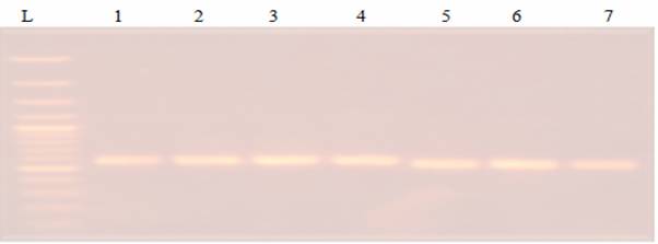
The extracted DNA from selected isolates of fungal specieswas successfully amplified with universal primer
ITS1 and ITS4 primer. The size of the amplicon of PCR
product was 1000bp (Figure 1). The analysis of the nucleotide sequences of ITS1
region of the isolates produced from their rDNA data
showed 7 identified isolates.
Figure 1: PCRAgarose gel
electrophoresis of genes amplified using ITS1 and ITS4 from DNA extracted of7 fungal
Isolates.
Thus, DNA from the isolates, which showed greater growth
in PDA, were extracted with good quality, and the PCR methodology applied with
the universal primers generated fragments of approximately 550 base pairs. With
the determination of the sequences of the amplified fragments and their
respective editions, and submitting the data to the analysis by BLAST, it was
found that the 7 isolates (Table 4) wereAlternaria
alternate, Aspergillusflavus,
A. fumigates, A. niger, Colletotrichumgloeosporioides, Fusariumverticillioides, Rhizopusstolonifer respectively.
Table 4: Molecular identification by
comparing the sequences obtained from the amplification of the genomic region
of the ITS of the rDNA using
BLAST in the NCBI
Amplicon No.
ITS 1 and ITS 4
SIMILARITY
SEQUENCE (NT)
1
Alternaria
alternate
100%
551
2
Aspergillusflavus
99%
641
3
Aspergillus
fumigates
99%
860
4
Aspergillusniger
100%
891
5
Colletotrichumgloeosporioides
99%
446
6
Fusariumverticillioides
99%
441
7
Rhizopusstolonifer
98%
398
DISCUSSION
Eleven fungal species were isolated from the infected
fruits, leaves and fruits of three varieties of Mangiferaindica. The isolated fungi were Alternariaalternata,
Aspergillusflavus,
A. fumigatus, A. niger, Colletotrichumgloeosporioides, Fusariumsemitectum, F. verticolor, Papulasporasp., Penicilliumsp., Pestalotiopsisguepiniiand Rhizopusstolonifer. All these isolates were identified as
responsible for the disease of mangoes in UsmanuDanfodiyo University, Sokoto.
This result is also consistent with reports by several
workers implicating C. gloeosporioidesas the
causal agent of diseases in mango (Than et al., 2008; Kim et al.,
2008; Sangeetha and Rawal,
2009; Jayasinghe and Fernando, 2009). This study
identified F. verticillioidesand L. theobromaetobe associated
with anthracnose of mango, implying that other fungal species may act in
synergy with C. gloeosporioidesto cause
fungal disease of mango. Several workers including Johnson (2008), had
implicated other fungal species in earlier reports, to be responsible for
postharvest diseases of mango associated with fruit rotting during ripening,
worldwide. Okerekeet al. (2010) also reported
the isolation of these fungal species from infected mangoes in their study.
Maqsoodet
al. (2014) identified C. gloeosporioides, Lasiodiplodiatheobromae, Alternariaalternata, Aspergillusnigerand Dothiorelladomonicanafrom Sindhri mango fruits, in which C. gloeosporioideswas found to be the most prevalent in
Pakistan. Rajmane and Korekar
(2016) reported that Alternariaalternata, Aspergillusflavus, A. fumigatus, A. niger, Botryodiplodiatheobromae, C. gloeosporioides,
Penicilliumchrysogenumand
R. stoloniferassociated with the spoilage of
mango fruit in India. Abdullah et al. (2016) found that Alternariaalternata,
A. aculeatus, Aspergillusflavus, A. japonicas, A. niger,
A. parasiticus, Eurotiumamstelodami, Mucorcircinelloides, Penicilliumviridicatum, Rhizopusarrhizus, Trichodermakoningii, T. harzianumand Verticilliumtenerumwere
associated with post harvest rot disease of mango in
Yemen. All the fungi associated with mango as pathogens deteriorate fruits.
In this study, a fragment was amplified by PCR from
fungal isolates with universal primer specific for distinguishing fungal
species. The nucleotide sequences of the ITS1 region of species, implying that
all the Colletotrichumspecies isolated
were C. gloeosporioidesand not C. acutatum. This result agrees with Nigeria Plant
Quarantine Service (2002) which listed C. gloeosporioidesas the causal agent of mango fruit anthracnose in Nigeria although there
were no representation of pathogen isolation and identification in their study.
For the identification of all the 7 fungal isolates that belonged to the 4genera, pathogenicity test that
confirm the pathogenesis of C. gloeosporioidesimplied
that, C. gloeosporioideswas the pathogen
responsible for anthracnose inmango, in Nigeria and
not C. acutatumas listed by CAB International
(2007).
CONCLUSION
Fungal diseases of mango have become a menace to many garden
and orchards in the study area, sometimes resulting in complete loss of the
crop in some locations. This identification method clearly identified the 7
most frequent fungal species isolated from mango fruits, leaves and stems in UsmanuDanfodiyo University, Sokoto. In addition, A. niger and C.
gloeosporioideswere found to be the fungus
responsible for anthracnose of mango in the study area.
ACKNOWLEDGEMENT
We wish to express our sincere gratitude to UsmanuDanfodiyo University, Sokoto and Tertiary Education Trust Fund (TETFUND, Abuja)
for sponsorship of this research.
CONFLICT OF INTEREST
The authors have declared that no competing interests
exist.
REFERENCES
Abdullah Q,
A Mahmoud and A Al-Harethi (2016).
Isolation and identification of fungal postharvest rot of
some fruits in Yemen. PSM Microbiology 1(1): 36-44.
Agrios, G.N
(2005). Plant Pathology.Fifth (5th)
edition. Pp. 27-557.
Barnett, H.L. and Hunter, B.B. (1998).Illustrated Genera of Imperfect Fungi.APS Press Pp.163-193.
CAB
International (2007). Pests excluded from Phytosanitary
measure assessments because they already occur in Nigeria. Retrieved
December 13, 2008, from CABI Crop Protection Compendium software.
Derso E. (1999):
Occurrence, prevalence and control methods of phaeoramularia
leaf and fruit spot disease of citrus in Ethiopia. Fruits, 54: 225–232.
Diedhiou, P.M., Mbaye, N. Drame, A. & Samb, P.I. (2007). Alteration of post-harvest
diseases of mango Mangiferaindicathrough production practices and climatic
factors.African Journal of Biotechnology, 6(9): 1087 -1094.
Etebu, E. & Osborn, A. M. (2012).Molecular approach to plant fungal
identification in agricultural soils.Nigerian
Journal Plant Protection, 26: 18 -34.
Hall, T.A. (1999).
BioEdit: A User-friendly biological sequence
alignment editor and analysis program for window 95/98/NT, Nucl.
Acids Symp. Ser. 41: 95 –
98.
Jayasinghe, C.K. & Fernando, T.H.P.S. (2009).First Report of Colletotrichumacutatumon Mangiferaindicain Sri Lanka.Cey.Jour. Sci. (Bio. Sci.)38(1): 31 – 34.
Johnson,
G.I. (2008). Status of mango post harvest disease
management R and D: Options and solutions for the Australian mango industry. Horticulture Australia final report for project MG08017.
Horticulture Australia p.130
Kim, J.K., Park, S., Choi, W., Lee, Y. & Kim, H.T. (2008).Characterization of ColletotrichumIsolates Causing Anthracnose
of Pepper in Korea.Plant Pathol.
Jour. 24(1): 17 – 23.
Kumar, A.S., Reddy, N.P.E., Reddy, K.H. & Devi, M.C.
(2007).Evaluation of Fungicidal
Resistance among ColletotrichumgloeosporioidesIsolates Causing Mango Anthracnose in Agri Export Zone of Andhra Pradesh, India.Plant Patho. Bull. 16:
157 – 160.
Maqsood A, A Rehman, I Ahmad, M Nafees, I
Ashraf, R Qureshi, M Jamil,
M Rafay and T Hussain (2014).
Physiological attributes of fungi associated with stem end rot of mango (MangiferaindicaL.)
varieties in postharvest fruit losses. Pak. J. Bot. 46(5): 1915-1920.
Nasiru A. M and Idris M (2021). Study of the Causes of Death of Mango Stands in Some Fadama Lands of Sokoto
Metropolis, Nigeria. Cross Current Int J Agri Vet Sci, 3(3), 15-21.
Nasiru, A. M. Salau, I. A and M. Yakubu, (2015), Fungi
Associated With Spoilage of Citrus Sinensisin
Fruits and Vegetables Market, Sokoto, Nigeria. Global Advanced Research Journal of
Agricultural Science 4(12): 919-922.
Nigeria Plant Quarantine Service (2001).Check-List of Plant Pests in Nigeria.NPQS Publ. Ibadan Univ. Printry,
Ibadan, Nigeria.
Okereke, V.C., Godwin-Egein, M.I.
& Arinze, A.E. (2010).Assessment of Postharvest Rot of mango
at Different Stages of market in Port Harcourt, Nigeria.Int’l Jour. Curr. Res.,11:006
– 010.
Okibo R.N.
(2001): Occurrence, pathogenicity and survival of Macrophomamangiferaein leaves branches and stems of mango
(MangiferaindicaL.). Plant Protect. Sci., 37: 138–144.
Rajmane SD and SL Korekar (2016). Isolation and identification of fungi associated with
spoilage of mango fruit, India. International Journal of Scientific Research
5(8): 2277-8179.
Sangeetha, C.G. & Rawal, R.D. (2009). Temperature Requirement of Different Isolates of ColletotrichumgloeosporioidesIsolated from Mango. American-Eurasian Journal
of Scientific Resources 4(1): 20 – 25.
Cite this Article: Abubakar,
MN; Salau, IA; Tambari, S
(2023). Fungal Pathogens Associated with Mango Diseases in UsmanuDanfodiyo University, Sokoto State, Nigeria. Greener Journal of Agricultural Sciences, 13(2): 163-169.