Greener Journal of Biomedical and Health Sciences
Vol. 9(1), pp. 87-96, 2026
ISSN: 2672-4529
Copyright ©2026, Creative Commons Attribution 4.0 International.

|
Greener Journal of Biomedical and Health Sciences Vol. 9(1), pp. 87-96, 2026 ISSN: 2672-4529 Copyright ©2026, Creative Commons Attribution 4.0 International. |
|
Hepatic Effects of Cigarette Smoke Exposure: Enzymatic and Histological Evidence from Wistar Rats
Chukwudi Francis Afuberoh¹*, Onyeka Chukwudi Okonkwo¹, Darlington Victor Nweze1, Anyaogu Charles Chinemeze², Okoye Ogochukwu Fidelis²
¹Department of Human Physiology, Faculty of Basic Medical Sciences, College of Health Sciences, Nnamdi Azikiwe University, Nnewi Campus, PMB 5001, Anambra, Nigeria.
²Department of Human Physiology, Faculty of Basic Medical Sciences, Chukwuemeka Odumegwu Ojukwu University, Uli Campus.
|
ARTICLE’S INFO |
|||
|
Article No.: 050726064 Type: Research Full Text: PDF, PHP, HTML, EPUB, MP3 DOI: 10.15580/gjbhs.2026.1.050726064
Accepted: 12/05/2026 Published: 16/05/2026
Keywords: Cigarette smoke exposure, Liver enzymes, Wistar rats, Hepatic histology, Hepatotoxicity
|
*Corresponding Author
Chukwudi Francis Afuberoh
Department of Human Physiology, Faculty of Basic Medical Sciences, College of Health Sciences, Nnamdi Azikiwe University, Nnewi Campus, PMB 5001, Anambra, Nigeria.
E-mail: fc.afuberoh@unizik.edu.ng
Phone: +2348039849331
|
Article’s QR code
|
|
|
|
|
|
|
|
ABSTRACT |
|
|
|
Cigarette smoke contains numerous toxic compounds capable of inducing systemic effects, including alterations in hepatic function. This study investigated the effects of chronic cigarette smoke exposure on liver enzyme activity and histological architecture in male Wistar rats. Twenty rats were randomly divided into two groups (n = 10 each): a control group and a smoke-exposed group. The exposed group was subjected to cigarette smoke inhalation daily for a period of four weeks, while the control group was maintained under normal laboratory conditions. At the end of the exposure period, blood samples were collected for biochemical analysis of liver enzymes, including alanine aminotransferase (ALT), aspartate aminotransferase (AST), and alkaline phosphatase (ALP). Liver tissues were also harvested for histopathological examination using standard staining techniques. Statistical analysis was performed using one-way analysis of variance (ANOVA), with significance set at p < 0.05. Results showed a significant decrease (p < 0.05) in serum ALT, AST, and ALP levels in the smoke-exposed group compared to the control. Histological evaluation revealed largely preserved hepatic architecture, with only mild inflammatory changes observed in some samples. The observed reduction in liver enzyme activity may suggest an alteration in hepatic metabolic function or possible enzyme suppression following chronic smoke exposure. In conclusion, chronic exposure to cigarette smoke modulates liver enzyme activity in Wistar rats, with minimal observable histological damage under the conditions of this study. Further investigations incorporating oxidative stress and molecular biomarkers are recommended to elucidate the underlying mechanisms.
|
|
|
|
INTRODUCTION
Cigarette smoking remains a leading cause of preventable disease and mortality worldwide, contributing significantly to the global burden of non-communicable diseases. Tobacco smoke contains thousands of chemical constituents, including nicotine, carbon monoxide, polycyclic aromatic hydrocarbons, and reactive oxygen species (ROS), many of which are known to exert toxic effects on biological systems [1,2]. While the deleterious effects of cigarette smoke on the respiratory and cardiovascular systems are well established, increasing evidence indicates that smoking also has systemic consequences, including adverse effects on liver function [3].
The liver plays a central role in the metabolism and detoxification of xenobiotics, making it particularly susceptible to damage from toxic compounds present in cigarette smoke. Exposure to these substances can induce oxidative stress, inflammation, and lipid peroxidation, which may compromise hepatocellular integrity and function [4,5]. Oxidative stress, in particular, has been identified as a key mechanism underlying smoke-induced tissue injury, resulting from an imbalance between the generation of reactive oxygen species and the antioxidant defense system, as demonstrated in experimental models of chronic cigarette smoke exposure [6, 7].”
Biochemical markers such as alanine aminotransferase (ALT), aspartate aminotransferase (AST), and alkaline phosphatase (ALP) are widely used indicators of liver function and hepatocellular injury. Elevations in these enzymes are commonly associated with membrane damage and leakage of intracellular contents into circulation [8]. However, experimental findings have shown variability in enzyme responses to toxic exposures, suggesting that factors such as duration, dose, and metabolic adaptation may influence hepatic enzyme activity [9].
Animal models, particularly Wistar rats, are frequently used to investigate the systemic effects of cigarette smoke due to their physiological and metabolic similarities to humans. Previous studies have demonstrated that chronic cigarette smoke exposure can lead to hepatic alterations, including changes in enzyme activity and histopathological features such as inflammatory cell infiltration, hepatocellular degeneration, and structural disorganization [10,11]. Additionally, cigarette smoke has been shown to exacerbate underlying liver conditions and contribute to the progression of liver diseases such as non-alcoholic fatty liver disease [3].
Histopathological examination remains an essential tool for evaluating liver injury, as it provides direct evidence of structural and cellular changes that may not be fully captured by biochemical markers alone. The combination of biochemical and histological assessments therefore offers a more comprehensive understanding of hepatic responses to toxic insults [12].
Despite existing evidence, inconsistencies in reported biochemical outcomes and variations in experimental conditions highlight the need for further investigation into the effects of chronic cigarette smoke exposure on liver function. This study was therefore designed to evaluate the impact of chronic cigarette smoke exposure on liver enzyme activity and hepatic histological architecture in male Wistar rats.
MATERIALS AND METHODS
Ethical Approval
All experimental procedures involving animals were conducted in accordance with internationally accepted guidelines for the care and use of laboratory animals. Ethical approval for this study was obtained from the Institutional Animal Ethics Committee (IAEC) of Nnamdi Azikiwe University. All efforts were made to minimize animal suffering and to reduce the number of animals used.
Study Location
This study was carried out in the Department of Physiology, Nnamdi Azikiwe University, Nnewi Campus. The experimental procedures, including animal handling, cigarette smoke exposure, and biochemical analyses, were conducted in the departmental laboratory. Histopathological examination of liver tissues was performed in the Department of Anatomy of the same institution.
Materials Used
Forty (40) male Wistar rats, Packs of commercial cigarettes (All stars),electronic weighing balance, Plain Blood tube (Fantastik, China), Distilled water, Standard Plastic Cages and water can, Latex Medical Hand gloves, Chloroform (Guondghuo, China), Top-Pro feed grower (JOS, Nigeria), Dissecting kits, Lighter, Plastic rectangular transparent container: Length (30m), Width (25m), (43m) wide, Cotton wool, plain bottles, Normal saline.
Experimental Animals and Housing
Forty (40) male Wistar rats with weight of 140 to 220 gram were obtained from the Animal House, Department of Physiology, Faculty of Basic Medical Sciences, College of Health Sciences, Nnamdi Azikiwe University, Nnewi Campus. Animals were kept in standard cages at a room temperature of 27±2°C. The animals were maintained with normal laboratory chow (Grower feed) and water ad libitium. Following acclimatization period of two weeks, the animals would be exposed to commercial cigarette smoke as the drug used in the study.
Experimental Design
Forty (40) male Wistar rats were randomly grouped into four groups of 10 animals each as follows:
Group A received rat chow and distilled water and libitum only
Group B were exposed to six sticks of cigarettes for 30 mins
Group C were exposed to six sticks of cigarettes for 60 mins
Group D were exposed to six sticks of cigarettes for 120 mins
The exposure to commercial cigarette smoke was done through whole body/chamber exposure and was administered once daily for 28 days between the hours of 7 am and 11 am.
Sacrifice and Sample Collection
Following 28 days of cigarette smoke exposure, the experimental animals were fasted overnight and sacrificed on day 30. The animals were euthanized under light anesthesia using chloroform in a closed chamber.
Blood samples were collected immediately after sacrifice via ocular puncture using heparinized capillary tubes and transferred into plain sample bottles. The collected blood was allowed to clot at room temperature for approximately 10–15 minutes and subsequently centrifuged at 3000 rpm for 10 minutes to obtain serum. The separated serum was carefully collected using a micropipette and stored for the determination of liver function parameters, including alanine aminotransferase (ALT), aspartate aminotransferase (AST), and alkaline phosphatase (ALP).
Following blood collection, the animals were dissected through a midline abdominal incision. The liver was carefully excised, rinsed in normal saline to remove blood stains, and blotted dry. The liver tissues were then fixed in 10% buffered formalin and preserved in labeled containers for histopathological examination.
Acute Toxicity Study of Cigarette Smoke
An acute toxicity study of cigarette smoke exposure was conducted in the Department of Physiology, Faculty of Basic Medical Sciences, College of Health Sciences, Nnamdi Azikiwe University, Nnewi Campus.
A total of forty (40) Wistar rats were used for this study. The animals were exposed to cigarette smoke via inhalation in a controlled chamber. The exposure protocol was adapted from the method described by Lorke (1983), with modifications to suit inhalational exposure to cigarette smoke rather than oral administration.
The animals were observed for signs of acute toxicity, including behavioral changes, respiratory distress, and mortality within 24 hours post-exposure.
Blood Collection and Termination of Experiment
At the end of the experimental period, the animals were fasted overnight and humanely euthanized under light chloroform anesthesia. Blood samples were collected via ocular puncture as described by Parasuraman et al. (2010) into plain tubes.
The blood samples were allowed to clot and then centrifuged at 3000 rpm for 10 minutes. The serum obtained was used for biochemical analysis of liver enzymes.
The liver was harvested through an abdominal incision, weighed, and rinsed in normal saline. The tissues were then fixed in 10% formal-saline and stored in airtight containers for subsequent histopathological processing and analysis.
Biochemical Analysis of Liver Function Parameters
Alanine Aminotransferase (ALT) Assay
Principle:
Alanine aminotransferase (ALT) catalyzes the transfer of an amino group from alanine to α-ketoglutarate, resulting in the formation of pyruvate and glutamate. The activity of ALT is determined spectrophotometrically based on the formation of pyruvate, as described by Reitman and Frankel (1957).
Procedure:
Serum ALT activity was determined using a commercially available diagnostic kit (e.g., Sigma-Aldrich, USA) following the manufacturer’s instructions. The absorbance was measured spectrophotometrically at the specified wavelength, and enzyme activity was expressed in units per liter (U/L).
Aspartate Aminotransferase (AST) Assay
Principle:
Aspartate aminotransferase (AST) catalyzes the transfer of an amino group from aspartate to α-ketoglutarate, forming oxaloacetate and glutamate. The enzyme activity is measured spectrophotometrically according to the method of Reitman and Frankel (1957).
Procedure:
Serum AST activity was measured using a standard commercial assay kit (e.g., Sigma-Aldrich, USA) in accordance with the manufacturer’s protocol. Absorbance readings were taken using a spectrophotometer, and results were expressed in U/L.
Alkaline Phosphatase (ALP) Assay
Principle:
Alkaline phosphatase (ALP) catalyzes the hydrolysis of phosphate esters in an alkaline medium, producing inorganic phosphate and alcohol. The activity is measured spectrophotometrically based on the rate of product formation.
Procedure:
Serum ALP activity was determined using a commercial diagnostic kit according to the manufacturer’s instructions. The absorbance was read at the appropriate wavelength, and enzyme activity was expressed in U/L.
Histological Procedure of the Liver
Fixation and Embedding
Liver tissue samples were fixed in 10% buffered formalin for at least 24 hours to preserve tissue architecture. The fixed tissues were then dehydrated in graded alcohol, cleared in xylene, and embedded in paraffin wax.
Sectioning and Staining
Paraffin-embedded tissues were sectioned at a thickness of 4–5 µm using a microtome. The sections were mounted on glass slides and stained with hematoxylin and eosin (H&E) following standard procedures as described by Bancroft and Gamble (2008).
Histopathological Examination
The stained sections were examined under a light microscope for structural and cellular changes, including hepatocellular degeneration, inflammatory cell infiltration, necrosis, and architectural distortion.
Statistical analysis
Data were analysed using one-way analysis of variance (ANOVA) followed by least significant difference (LSD) post-hoc test. Results were expressed as mean ± standard error of mean (SEM). Statistical significance was set at p < 0.05.
RESULTS
Table 1: Effect of Cigarette Smoke Exposure on Body Weight in Experimental Rats
|
|
Initial weight (g) |
Final weight (g) |
p-value |
t-value |
|
|
MEAN |
MEAN |
|
|
|
Group A (control) |
118.00 |
121.00 |
0.776 |
0.325 |
|
Group B |
147.30 |
190.30 |
0.268 |
1.286 |
|
Group C |
148.00 |
180.67 |
0.107 |
2.075 |
|
Group D |
164.33 |
193.00 |
0.212 |
1.483 |
Data were analyzed using paired Student’s t-test (GraphPad Prism version 9.5.1). Values were considered statistically significant at p < 0.05.
There was an observable increase in body weight across all experimental groups following the exposure period. However, the changes in body weight were not statistically significant in both the control and cigarette smoke-exposed groups (p > 0.05). This suggests that chronic cigarette smoke exposure did not significantly affect body weight gain in the experimental rats during the study period.
Table 2: Effect of Cigarette Smoke Exposure on Relative Liver Weight in Experimental Rats
|
|
Rel. liver weight (g) |
|
|
MEAN |
|
Group A (control) |
3.11 |
|
Group B |
2.66 |
|
Group C |
2.42 |
|
Group D |
3.44 |
|
F-ratio |
1.918 |
Data were analyzed using one-way analysis of variance (ANOVA) followed by Least Significant Difference (LSD) post hoc test. Values were considered statistically significant at p < 0.05.
Table 2 shows the effect of cigarette smoke exposure on relative liver weight in experimental rats. There was a decrease in the mean relative liver weight in Groups B and C compared to the control group (Group A), while Group D showed a slight increase. However, these differences were not statistically significant (p > 0.05), as indicated by the ANOVA result (F = 1.918). This suggests that cigarette smoke exposure did not significantly affect relative liver weight in the experimental rats.
Table 3: Effect of Cigarette Smoke Exposure on Liver Enzymes in Experimental Rats
|
|
AST level (IU/L) |
ALT level (IU/L) |
ALP level (IU/L) |
|
|
MEAN |
MEAN |
MEAN |
|
Group A (control) |
118.56 |
80.63 |
120.59 |
|
Group B |
95.63 |
73.42 |
112.22 |
|
Group C |
89.17 |
73.77 |
101.72 |
|
Group D |
92.18 |
72.77 |
111.19 |
|
F-ratio |
34.19 |
6.779 |
10.51 |
Data were analyzed using one-way analysis of variance (ANOVA) followed by Least Significant Difference (LSD) post hoc test. Values were considered statistically significant at p < 0.05.
* indicates significant difference compared to control group.
Table 3 shows the effect of cigarette smoke exposure on liver enzyme activities in experimental rats. There was a statistically significant decrease (p < 0.05) in serum AST levels in Groups B, C, and D when compared to the control group (Group A). Similarly, serum ALT levels were significantly reduced in all smoke-exposed groups relative to the control.
In addition, ALP activity was significantly decreased in Groups B, C, and D compared to Group A. The ANOVA results (AST: F = 34.19; ALT: F = 6.779; ALP: F = 10.51) indicate that these differences across groups were statistically significant.
Overall, chronic cigarette smoke exposure resulted in a significant reduction in liver enzyme activities in the experimental rats.
HISTOLOGICAL FINDINGS
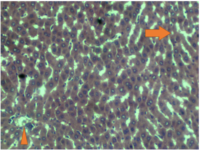
Plate 1 (Group A – Control)
Photomicrograph of liver tissue from the control group showing normal hepatic architecture. The section reveals well-organized hepatic lobules with radiating cords of hepatocytes (arrow) and sinusoids lined by intact endothelial cells (arrowhead). The hepatocytes appear polygonal with centrally located nuclei and eosinophilic cytoplasm.
(Hematoxylin and eosin stain, ×400)
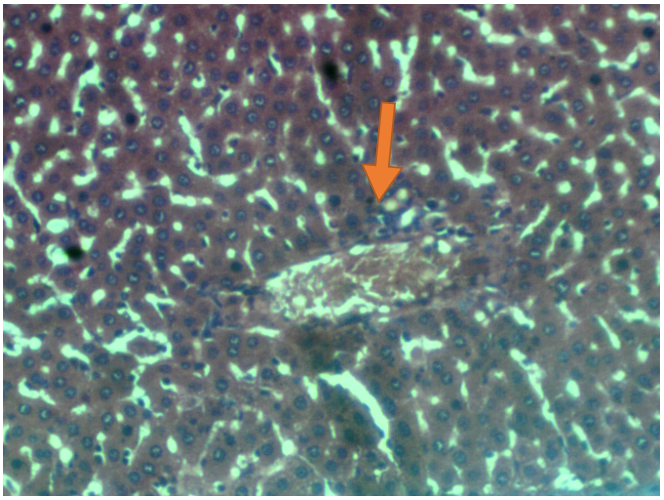
Plate 2 (Group B – Smoke-Exposed)
Photomicrograph of liver tissue showing mild inflammatory changes, characterized by slight infiltration of inflammatory cells around the central vein (arrowhead). The overall hepatic architecture is largely preserved.
(Hematoxylin and eosin stain, ×400)
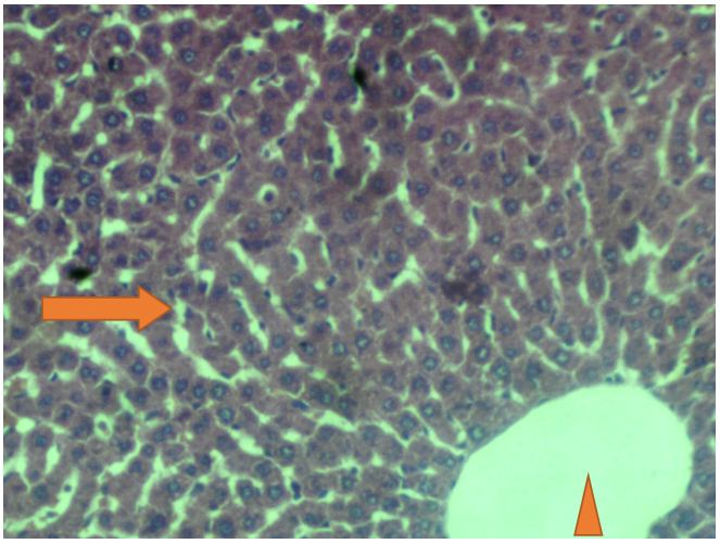
Plate 3 (Group C – Smoke-Exposed)
Photomicrograph of liver tissue showing largely normal hepatic architecture with well-organized hepatic cords (arrow). The central vein appears mildly dilated with intact endothelial lining (arrowhead). Hepatocytes retain normal morphology with prominent nuclei and eosinophilic cytoplasm.
(Hematoxylin and eosin stain, ×400)
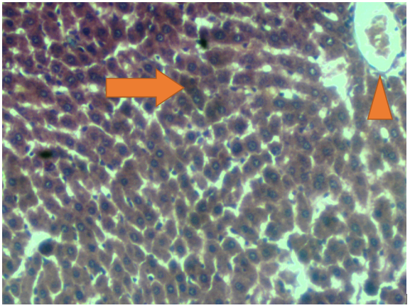
Plate 4 (Group D): Photomicrograph of a section of liver tissue of the albino rat shows normal histological features, including well-organised hepatic lobules with radiating hepatocyte cords (arrow) and central lined by continuous endothelial cells (arrowhead). The hepatocytes are polygonal with prominent nuclei and eosinophilic cytoplasm (Staine with H&E X400)
DISCUSSION
The present study investigated the effects of chronic cigarette smoke exposure on liver enzyme activity and histological architecture in male Wistar rats. The findings revealed a significant decrease in serum liver enzymes (AST, ALT, and ALP) in smoke-exposed groups compared to the control, while histological examination showed largely preserved hepatic architecture with only mild inflammatory changes.
Cigarette smoke contains numerous toxic constituents, including reactive oxygen species (ROS), nicotine, and polycyclic aromatic hydrocarbons, which have been widely reported to induce oxidative stress and tissue injury in various organs, including the liver [1,2]. The liver, being the primary site for detoxification and metabolism of xenobiotics, is particularly susceptible to damage from such toxic exposures [3]. Previous studies have commonly reported elevated liver enzyme levels following cigarette smoke exposure, indicative of hepatocellular injury and membrane damage [4,5].
However, in contrast to these findings, the present study demonstrated a significant reduction in AST, ALT, and ALP levels in all smoke-exposed groups. This observation, although less commonly reported, is not entirely unprecedented. Some studies have suggested that chronic exposure to toxic agents may lead to suppression of hepatic enzyme synthesis or altered enzyme activity due to adaptive metabolic responses [6]. Additionally, prolonged exposure to cigarette smoke may impair hepatocellular function at the enzymatic level without causing overt structural damage, resulting in reduced enzyme release into circulation [8].
Another possible explanation for the observed decrease in enzyme levels is the inhibition of enzyme activity by toxic constituents of cigarette smoke. Nicotine and other compounds have been shown to interfere with normal cellular metabolism and enzymatic processes, potentially leading to decreased measurable enzyme activity in serum [9]. Furthermore, oxidative stress induced by cigarette smoke may alter protein structure and function, thereby affecting enzyme production and release, as demonstrated in experimental models showing increased lipid peroxidation and reduced antioxidant activity following chronic smoke exposure [7,10].
The histopathological findings of this study support the biochemical results. Liver sections from smoke-exposed groups showed largely normal hepatic architecture, with only mild inflammatory infiltration observed in Group B. There was no evidence of severe hepatocellular degeneration, necrosis, or fibrosis. This suggests that, under the conditions of this study, cigarette smoke exposure did not produce significant structural liver damage. Similar findings have been reported in some experimental studies where mild or no histological alterations were observed despite biochemical changes, with preservation of tissue architecture despite underlying oxidative stress [11, 7].
The absence of marked histopathological damage alongside reduced enzyme levels may indicate an early or adaptive phase of hepatic response to cigarette smoke exposure. It is possible that the duration or intensity of exposure in this study was insufficient to induce severe liver injury. Alternatively, the liver may have activated compensatory mechanisms to maintain structural integrity despite biochemical alterations [12].
The findings of this study therefore suggest that chronic cigarette smoke exposure may modulate liver enzyme activity without causing significant histological damage within the exposure period. This highlights the complexity of hepatic responses to toxic insults and underscores the importance of combining biochemical and histological assessments in evaluating organ toxicity.
However, this study has some limitations. The absence of oxidative stress markers and molecular indicators limits the ability to fully elucidate the mechanisms underlying the observed changes. Future studies incorporating parameters such as lipid peroxidation, antioxidant enzyme activity, and inflammatory cytokines would provide deeper insight into the effects of cigarette smoke on liver function.
CONCLUSION
In conclusion, this study demonstrated that chronic cigarette smoke exposure significantly reduced serum liver enzyme activities (AST, ALT, and ALP) in male Wistar rats, without causing marked histopathological alterations in liver tissue. The observed decrease in enzyme levels, alongside largely preserved hepatic architecture, suggests that cigarette smoke may induce functional changes in liver metabolism rather than overt structural damage under the conditions of this study.
These findings indicate that biochemical alterations may occur in the early or adaptive phase of exposure prior to the development of severe hepatic injury. However, the absence of oxidative stress and molecular markers limits the ability to fully elucidate the underlying mechanisms. Further studies incorporating oxidative stress parameters, inflammatory markers, and longer exposure durations are recommended to better understand the hepatic effects of chronic cigarette smoke exposure.
Declarations and Acknowledgement
The authors acknowledge the technical staff of the Department of Human Physiology, Nnamdi Azikiwe University, Nnewi Campus, for their assistance during the experimental procedures.
Funding/Sponsorship
This research received no specific grant from any funding agency in the public, commercial or not-for-profit sectors.
Conflict of Interest
No conflict of interest associated with this work.
Contribution of Authors
We declare that this work was done by the authors named in this article and all liabilities pertaining to claims relating to the content of this article will be borne by the authors. UJC conceived and designed the study, carried out the experiments and drafted the manuscript. OOC supervised the study, analysed the data and revised the manuscript. All authors read and approved the final manuscript for publication.
Availability of Data and Materials
The datasets used and/or analysed during the current study are available from the corresponding author upon reasonable request.
Use of Artificial Intelligence/Large Language Models
Artificial intelligence-based tools were used for language editing and formatting of the manuscript only. No AI system was used for data generation, analysis or interpretation.
REFERENCES
Afuberoh FC, Umeji JC, Anyaogu CC, Okoye OF. Impacts of chronic cigarette smoke exposure on lung histology and oxidative stress markers in adult male Wistar rats. Eur J Biomed Pharm Sci. 2026;13(1):100–106. doi:10.5281/zenodo.18094443
Azzalini L, Ferrer E, Ramalho LN, Moreno M. Cigarette smoking exacerbates nonalcoholic fatty liver disease in rats. Hepatology. 2010;51(5):1567–1576. doi:10.1002/hep.23516
Azzalini L, Ferrer E, Ramalho LN, Moreno M. Smoking and liver disease progression. Hepatology. 2010;51(5):1567–1576. doi:10.1002/hep.23516
Bandiera S, Pulcinelli RR, Huf F, Almeida FB. Hepatic and renal damage induced by cigarette smoke in rats. Toxicol Res. 2021;10(2):321–330. doi:10.1093/toxres/tfaa093
Czekaj P, Pałasz A, Lebda-Wyborny T. Morphological changes in liver of rats exposed to cigarette smoke. Int Arch Occup Environ Health. 2002;75(Suppl):S27–S32. doi:10.1007/s00420-002-0343-3
El Golli N, Jrad-Lamine A, Neffati H. Impact of cigarette smoke on liver function in rats. Toxicol Mech Methods. 2016;26(7):1–9. doi:10.3109/15376516.2016.1160963
Kumar V, Abbas AK, Aster JC. Robbins and Cotran Pathologic Basis of Disease. 10th ed. Elsevier; 2020.
El-Sokkary GH, Cuzzocrea S, Reiter RJ. Effect of chronic nicotine administration on oxidative stress in rat liver. Toxicology. 2007;239(1–2):60–67. doi:10.1016/j.tox.2007.06.102
Giannini EG, Testa R, Savarino V. Liver enzyme alteration: a guide for clinicians. CMAJ. 2005;172(3):367–379. doi:10.1503/cmaj.1040752
Talhout R, Schulz T, Florek E, van Benthem J, Wester P, Opperhuizen A. Hazardous compounds in tobacco smoke. Int J Environ Res Public Health. 2011;8(2):613–628. doi:10.3390/ijerph8020613
Valko M, Rhodes CJ, Moncol J, Izakovic M, Mazur M. Free radicals and antioxidants in normal physiological functions and human disease. Int J Biochem Cell Biol. 2007;39(1):44–84. doi:10.1016/j.biocel.2006.07.001
World Health Organization. WHO report on the global tobacco epidemic 2021. Geneva: WHO; 2021. Available from: https://www.who.int
|
Cite this Article: Afuberoh, CF; Okonkwo, OC; Nweze, DV; Anyaogu, CC; Okoye, OF (2026). Hepatic Effects of Cigarette Smoke Exposure: Enzymatic and Histological Evidence from Wistar Rats. Greener Journal of Biomedical and Health Sciences, 9(1): 87-96, https://doi.org/10.15580/gjbhs.2026.1.050726064. |