Nweke, EO; Elemuo, S; Usige, E; Offor, C; Igwedibia, P; Ifedi, I; Okeke, JC; Oguwike, FN; Oguwike RC; Oguwike, ER (2022).
|
Greener
Journal of Medical Sciences
Vol. 12(1),
pp. 91-95, 2022
ISSN:
2276-7797
Copyright
©2022, the copyright of this article is retained by the author(s)
https://gjournals.org/GJMS
|

|
Histological and Haemostatic
Effect of Aqueous Extract of Lycopersicon esculentum (Tomatoes) On Wound Healing and Liver Cells
of Male Albino Wistar Rats.
Nweke, E.O1; Elemuo,
S1; Usige, E1; Offor, C2; Igwedibia,
P3; Ifedi, I3; Okeke, J.C3; Oguwike,
F.N3; Oguwike, R.C4; Oguwike, E.R5
1.
Dept. of
Anatomy, Faculty of Basic Medical Sciences. Chukwuemeka
Odumegwu, Ojukwu University
Uli campus, Anambra state.
2.
Dept. of
Medical Biochemistry, Faculty of Basic Medical Laboratory Science. Chukwuemeka Odumegwu, Ojukwu University Uli campus, Anambra state.
3.
Dept. of
Physiology, Faculty of Basic Medical Sciences. Chukwuemeka
Odumegwu, Ojukwu University
Uli campus, Anambra state.
4.
Dept. of
Pharmacology, Faculty of Pharmaceutical Sciences. Chukwuemeka
Odumegwu, Ojukwu University
Igbariam campus, Anambra
state.
5.
Dept. of
Medicine and Surgery, College of Medicine. Chukwuemeka
Odumegwu, Ojukwu University
teaching Hospital Amaku- Awka.
Anambra state Nigeria.
|
ARTICLE INFO
|
ABSTRACT
|
|
Article No.:021422019
Type: Research
Full Text: PDF, HTML, EPUB, PHP
|
It is often
believed that most plants in ethno-medical practices possess medical values, hence for thousands of years, people have resorted
to medicinal plants as source of healing. The histological and haemostatic
effect of aqueous extract of Lycopersicon esculentum (Tomato) on wound healing and the liver
cells of male albino wistar rats was investigated
using seventy (70) rats weighing 150-180g. The control group was fed normal
rat feed and drinking water ad libitum while the test rats were given normal
rat feed and water ad libitum in addition to administration of 1.0ml once
daily of the fruit extract of Lycopersicon esculentum. Blood samples were obtained from the
tails of the animals into EDTA bottles and sodium citrate bottles for
analysis, later the animals were stunned and their liver extracted and placed
in formalin for tissue processing and examination. The blood samples were
examined for haematological and haemostatic test. Result indicate that the
extract of Lycopersicon esculentum
has no significant effect on haemoglobin concentration, packed cell volume
and white blood cell count, while there was a significant effect on the
platelet count (P<0.01), Bleeding and Clotting time (P<0.05) resulting
to shortening of their time. The histological result showed no distortion in
the cyto- architecture of the livercells.
It could be deduced from this work that Lycopersicon esculentum
contains biological principle that can affect haemostasis.
|
|
Accepted: 17/02/2022
Published: 25/03/2022
|
|
*Corresponding Author
Prof. F.N Oguwike
E-mail: foguwike@ gmail. com
Phone: 08037791363
|
|
Keywords: Haemostasis, Lycopersicon esculentum,
Histology, Prothrombin time, bleeding time, wound
healing, liver cells.
|
|
|
|
INTRODUCTION
Crude extracts of some plants could play roles in hastening the haemostatic activities in a damaged tissue when applied
topically on the wound or cut to arrest bleeding and hasten healing of wound.
Such plant is Lycopersicon esculentum
(Tomato). This study is to know the histological and haemostatic
effect of aqueous extract of Lycopersicon esculentum on wound healing and liver cells of male
albino wistar rats.
Wound healing is the process of repair following injury to the skin and
other soft tissues. Initial stages of wound healing involve an acute
inflammatory phase followed by synthesis of collagen and other extracellular
matrix which are later remolded to for a scar (Ligha
et al, 2008).
Wound healing is influenced by many factors including the kind of
medicine used to accelerate the wound healing process and to prevent infection
(Prockop et al, 1995). The healing response is
characterized by the movement of specialized cells into the wound site.
Platelets and inflammatory cells are the first to arrive at the site of injury
and they provide key functions and “signals” needed for the influx of
connective tissue cells and a new blood supply. These chemical signals are
known as cytokines or growth factors (Lawrence et al, 1994).
Lycopersicon esculentum belongs
to the same family with eggplant and potato, solanacae
(Ogundipe 2004). It is one of the most widely grown
vegetable crop in the world and also one of the common
vegetables grown in home gardens and in every garden in Nigeria.
Chemical contents and Medicinal value:
Lycopersicon esculentum
contains calories, protein, carbohydrates, fat, sodium, potassium, dietary
fiber, vitamin A (20%), vitamin C (40%), potassium
(10%), Iron (2%) (Peralta et al, 2001). They are rich in calcium and phosphorus.
Being rich source of vitamin A, Lycopersicon esculentum are dependably preventive against night
blindness, short sightedness and other diseases of the eye.
The liver is the largest organ in the body (Baker et al, 1998) and from
metabolic stand point is the most complex. Tests of its many functions have
been devised in the hope that they will serve as diagnostic aids when a
metabolic process has been disturbed. The adult liver weighs about 1500g is
located beneath the diaphragm. It produces coagulation factors.
Homeostasis is a process of arrest of blood loss (Guyton, 2006).
Whenever a tissue is damaged, haemostatic activity occurs
by successive mechanisms which include vascular spasm, formation of blood clot,
platelet aggregation and growth of tissues into the blood clot.
MATERIALS AND METHODS
Selection of Animals:
For the haemostatic and histological studies, seventy (70) rats
were used, weighing 150-180g. Male rats were kept separately for 30 days
feeding.
Preparation of Extracts:
The method used
here is as described by Samuelson et al 1992.Fresh fruits Lycopersicon esculentum were collected, weighed,
washed, cut and blended ina
mortar with a pestle. Thereafter, 50mls of the extract was filtered out using Whatman No.1 filter paper. The extract was dried by
evaporation to dryness .0.5g of the dried extract was dissolved in 1000 ml of
the sterile water to give to the animals. They also received their normal rat
pellets and drinking water along with the control rats ad libitum.
·
Phytochemical analysis of the fruits
Toxicity study:
The LD50
of the extract in albino Wister rats was determined using Lorke’s
method. The procedure of determining the lethal dose is by increasing the
concentration of the extracts
administered to the rats (after weighing them) ineach
of the groups consisting of eight (8) rats per group for five (5) days.
The concentration
given was at the rate of 1000mg/kg, 1500mg/kg, 2000mg/kg, 2500 mg/kg, 3000mg/kgand 4000 mg/kg. The percentage rate of their death and survival
was noted and a graph was plotted to determine the LD50.
Haemostatic mechanism
test:
·
The haemoglobin, packed cell volume and WBC counts were done
using the method described by Baker et al, (1998).
·
Platelet count
estimated was done by the method of Brecher and
Cronkite (1950).
·
The whole blood
clotting time was estimated by the method of Lee and White.
·
The bleeding
time was carried out by the method of Dejana.
Histological studies:
The histological sections of the liver were stained with Eosin and haematoxylin (H&E staining) as described in Baker et al
1985. These studies were later examined by a histologist.
Phytochemical analysis of fruits was done using standard phytochemical
screening method described by Trease and Evans
(1985), Harban (1972).
Experimental Designs:
This study was divided into two (2) different studies namely: Study A
and Study B.
Study A: It is the oral
administration of the aqueous extracts of L.
esculentum to the test rats Group B (50), while
the control rats Group A (20) received rat feed. This
lasted for 30 days.
Study B: in this study, prothrombin time (PT)
and partial thromboplastin time with kaolin test
(PTTK) were done to establish the possible pathway of the action of the
extracts. The concentration of extracts used was 5mg/kg.
Collection of samples for study:
2.0ml of blood sample was collected from each rat in all the groups
(A&B) into EDTA bottle to determine the initial blood pictures before
feeding them on the extract. Their weight and full blood count (FBC) including
platelet count, bleeding time, clotting time, prothrombin
time and partial thromboplastin time kaolin were
monitored. At the end of 30 days of feeding on the extract, blood samples were
again obtained from the tails of the animals for the various tests as monitored
earlier. The liver of the test rats and control rats were extracted by stunning
the animals and using sterile blood lancets to remove them, they were kept in a
formalin container and later used for tissue processing and staining.
Statistical Analysis:
The result obtained in the study for haemoglobin
concentration (mg/dl), packed cell volume, WBC count, platelet counts, bleeding
time prothrombin time test, partial thromboplastin time kaolin and clotting time test were
represented as mean and standard deviation (Mean + S.D), while students-t-test
was used to compare the result of the control and tests a P value of less than
or equivalent to P<0.05 or P=0.05) was considered statistically significant.
Results
Table 1: Phytochemical study
|
|
Alkaloids
|
Glycosides.
|
Calcium.
|
Carbohydrate.
|
Flavonoids,
Steroids, Tannins, Fats & Oil, Saponins,
Terpenoids.
|
|
Degree of concentration
|
+++
|
++
|
+++
|
-
|
-
|
Key: - (Negative) + (Present in small concentration) ++ (Present in moderate concentration, +++ (Present in large concentration).
Table 2:Haematologicalindicies of albino wistar rats before the
administration of extracts and 30 days following the administration of L. esculentum.
|
Groups
|
Hbg/dl + S.D
|
PCV L/L + S.D
|
Platelets count * 109+
S.D
|
WBC count/mm3+ S.D
|
Bleeding time sec+ S.D
|
Clotting
time sec+
S.D
|
|
Group A (control) n=10, Extract free
|
12.3+ 0.4
|
36.9 +0.3
|
174 +
402
|
5,246 + 0.5
|
2.5 + 0.8
|
5.8 + 0.5
|
|
Day 1 before L.esculentum
|
12.0+ 0.5
|
36.0+ 0.5
|
168 + 315
|
5,830+ 0.3
|
2.7 + 0.5
|
5.7+ 0.6
|
|
30 days after administration of L.esculentum
Group B (50)
|
11.2 + 0.7
|
33.6+ 0.9
|
172+418
|
5,190+1.6
|
1.6 + 0.5
|
3.7 + 0.5
|
|
Significance
|
P>0.05
|
P>0.05
|
P>0.01
|
P>0.05
|
P<0.01
|
P<0.01
|
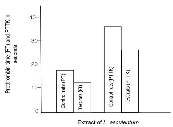
Figure 1: The effect of L.esculentum on
the prothrombin time and partial thromboplastin
time kaolin of albino wistar rat

Fig 2: Lethality studies
indicating the effects of administering graded doses (1,000mg/kg I.P rat) of
the fruit extract of L. esculentum (tomatoes) against the percentage
mortalities. LD50= 2,500mg/kg body weight.
DISCUSSION
The histological and haemastatic effect of
aqueous extract of Lycopersicon esculentum
(Tomatoes) on wound extract healing and liver cells of male albino wistar rats has been studied.
The result of the lethal studies showed that the LD50 in rats
using extract of L. esculentum
was 2,500mg/kg (Fig.2). The dose used in this study (5mg/kg) was far below the
lethal dose and so was considered safe to the animals used throughout the
period of study. This therefore shows that the result obtained with the plant
material was authentic and nontoxic. The phytochemical analysis of the extract
indicates the presence of typical plant constituents (Okoli
et al, 2007) such as alkaloids, calcium, saponins,
steroids, terpenoids, carbohydrates, glycosides,
tannins, acidic compounds in the extract.
The alkaloids (protein precipitants) and calcium contained in this plant
extract are known to be involved in the precipitation of coagulation factors (Taofeeq et al, 2005) for clotting to occur fast.
On red cell indices, there is no significant change in both the Hb concentration PCV in rats fed with the extracts of L. esculentum.
This agreed with the rich vitamin C, protein, and iron contents earlier
reported (Peralta et al, 2001). L. esculentum extract has no effect on white blood cell
count but has a little effect on increasing platelet count (Table 2). The
extract arrested bleeding time hence demonstrating haemostatic
activity. The prothrombin time and partial thromboplastin time were shortened by the extracts of L. esculentum.
The high concentration of calcium and alkaloids in L. esculentum may have contributed to its
ability in shortening bleeding and clotting time. On the histological results
of the state of the liver cells, it could be noticed that there was high
improvement of the performance of liver cells in the test rats than in the
control rats. There is no distortion of the cyto-architecture
of the liver cells. This tallies with the haematological
and haemostatic results hence the reduction in the
bleeding and clotting time of test rats.
The liver plays an important role in the formation and destruction of
red cells (Oguwike 2013). The liver also forms a
large portion of the bloodsubstances utilized in the
coagulation process. They are namely fibrinogen, prothrombin
accelerator, prothrombin, accelerator globulin
(factor V), factor VII and several other less important coagulation factors.
Vitamin K is required by the metabolic process of the liver for the formation
of prothrombin and factors VII. IX
and X. It could be deduced from this work that the presence of high
quantity of calcium and alkaloids in L. esculentum and high performance of liver cells in
producing coagulation factors during the intake of L. esculentum extracts could have
contributed to rapid healing of wounds in the rats.
REFERENCES
1.
Baker F.J, Silverton R.E (1985). Investigation for Haemostatic abnormalities.
Introduction to Medical Laboratory Technology p348. Butter worths
publication, Woburn London.
2.
Baker F.J, Silverton R.E, Palister C.J (1998). The liver function tests. Introduction to medical laboratory technology
7th edition p130. Printed by Linacre House Jordan Hill Oxford Ox2
8DP, 225 wildwood Avenue weburn Ma 01801-2041.
3.
BrecherL, and Cronkite M (1950).
Platelet count in Dacie J. V ed. Basic Haematological Techniques practical haematology
6th edition. Churchill Livingstone, Edinburgh London, Melbourne p43.
4.
Dejana E, Villa S, de Gaetano, (1982). Bleeding time in rats, a comparison of different experimental
condition. Thrombo Haemostato
48: 108-111.
5.
Guyton A. C, (2006). Haemostasis and Blood
coagulation. Textbook of Medical physiology 11th edition p457.
Published by Eiseveri, a division of Reed Elsevier.
Private united Sir, Pratap, Udyong
captain Guar Marg. Srini was puri
Delhi 110065 India.
6.
Harbourne J.B.C (1973). Phytochemical method. Chapman and Hall, London p273.
7.
Ligha A.E, Fawehimi H.B
(2008). Histological observations of the wound healing
properties of plant exudates of Jatropha curcas Linn. Journal of experimental and clinical
Anatomy 7(1) 17-21.
8.
Lawrence W.T, Diegelmann R.F (1994). Growth factors in wound healing. Clin. Dermatol 12:157.
9.
Lorke D, (1985). A new approach on practical acute toxicity testing. Arch Toxicity 54:
275-278.
10.
Ogundipe .O, Anyanuw K,
(2004): Tomatoe juice may cause clotting in diabetic rats with oral cancer. A newspaper repeat in Guardian newspaper Feb 24 p37.
11.
Oguwike F.N, (2013). Functions of the liver comprehensive Medical Physiology p98 Printed and
Published in Nigeria by Ideal way Publications Nigeria. 39 Nike Road Abakpa Nike Enugu Nigeria.
12.
Okoli C.O, Akah P.A, Okoli A.S (2007). Potentials of leaves of Asphilia Africana
(compositae) in wound care: an experimental
evaluation BMC complement. Allen Med. 2007: 7: 24 Published online 2007 July 10
Doi: 10, 1186/.
13.
Peralta I.E, Spooner D.M (2001). Granule bound starch synthetase. Lycopersicon (mill). American Journal of Botany 88 (10)
p1888-1902.
14.
Prockop D.J, Kivirriko K. J,
(1995). Collagen molecular biology, diseases and
potentials form therapy. Annual Rev. Biochem 64:403.
15.
Samuel G, Faraha K, Percleason
E, (1992). Inventory of plants used in Traditional
Medicine in Namibia. Plants of the families’ coubretacea.
Journal of Ethno-pharmacology p68-70.
16.
Taofeeq O, Oyeniyi M.A, Tesleem A.O (2005). Mechanism of action of Jastropha gossypifolia, a stem latex as
a haemostatic agent. European Journal of General
Medicine.
17.
Trease G.E, Evans W.C (1996). Textbook of pharmcognosy 14th ed.
W.B Saunders London p14.
|
Cite this Article: Nweke, EO; Elemuo,
S; Usige, E; Offor, C; Igwedibia, P; Ifedi, I; Okeke, JC; Oguwike, FN; Oguwike RC; Oguwike, ER (2022).
Histological and Haemostatic Effect
of Aqueous Extract of Lycopersicon Esculentum (Tomatoes)
On Wound Healing and Liver Cells of Male Albino Wistar
Rats. Greener Journal of Medical
Sciences, 12(1): 91-95.
|